Wednesday, March 25, 2020

WHY TELEMEDICINE?

Written by: Dr. Robert L. Bard

The communication features of TeleMed can also be used to
train other doctors in remote locations on new equipment
or modalities in patient care-
One of the current updates in patient care is the advancement of TELEMEDICINE. Thanks to the expansion of our web-based means of communication, doctors and patients are now able to convert the standard office visit to a virtual consultation or a remote, face-to-digital face exchange. Patients enjoy the benefits of the complete elimination of costly travel time and the potential viral exposure of sitting in a busy waiting room.

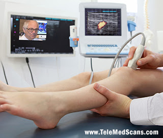
By definition, TeleMedicine is the implementing of any health-related services and information through the use of electronic and telecommunication technologies. It allows for REMOTE patient and clinician contact, care, advice, reminders, education, intervention, monitoring, and remote admissions.

Conceptually, TeleMed technologies are used as an alternative means of communication, resulting in a more efficient use of the doctors' and patients' time.  By communicating on a video-conferencing platform, TeleMedicine allows the physician to easily share files, records and imaging on the same screen that the doctor and patient are conducting a private consultation. TeleMed has been recognized to expand the practitioner's ability to reach more patients geographically and in less time- thus growing the medical practice this way.

To date, many new advancements have been added to this growing concept of remote patient care - including REAL-TIME INTERACTIVE SERVICES or "mHEALTH", REMOTE MONITORING (RPM), VIRTUAL CONSULTS (a form of live video conferencing) and STORE-AND-FORWARD (also known as 'asychronous video').Whether it's to reduce medical costs, save time or stay safe from potential (or advancing) health risks, methods of patient care continues to evolve toward better performance and efficiency for the convenience of the patient as well as improved use of time for the provider. This includes the smart use of internet technology and online private portals to offer a virtual alternative to the doctor's office visit.


PATIENTS WITH CHRONIC CONDITIONS & ALL IMMUNO-COMPROMISED ARE AT HIGH RISK

Chronic conditions like lung and heart disease, lupus, and diabetes directly affect the immune system. Also patients diagnosed with auto-immune disorders and cancers (including those currently undergoing chemotherapy treatments) can leave a person immuno-compromised. This makes them a very high risk of contracting deadly viruses like the Coronavirus. Anyone with weakened immune systems are highly advised to stay indoors and prevent any contact with the public. To battle this pandemic the right way, we all need a strong IMMUNE SYSTEM to recover from contamination. Also, ask any of your doctors if they subscribe to TeleMedicine as an alternative to any upcoming office visits.

THE FUTURE OF HOUSE CALLS: A SMART UPGRADE TO DOCTORS VISITS
Within the past several decades, the medical community has been put into overdrive to come up with new solutions (or modify existing ones) to implement safer, more efficient and cost-effective ways of working with the public. From the global demand for active medical personnel, to the rising wave of safety concerns that of the many at-risk patients, we face a great need to upgrade patient care.

Elderly patients or those with chronic conditions may find it difficult (and even precarious) to travel to their doctors' office and sit in a waiting room with other sick people with unknown conditions. Advancing this scenario to an electronic doctor's visit or TELEMEDICINE is an available reality that can easily address this concern. SO MUCH CAN BE DONE FROM HOME!

TELEMED BENEFITS FOR THE PATIENT
• Convenient & cost effective
• No Transportation needed
• No need for time off work or child care
• Eliminate the waiting room
• Quicker access to all specialists
• Improved access to files and prescriptions


Additional Articles (also see):
• Coronavirus Prompts Spike in TeleMedicine Use- By: Dr. Stephen Chagares
• Surviving a Pandemic with Perspective - By: Lennard Gettz
................................................................................................................................................................
CONTRIBUTING WRITER

ROBERT L. BARD, MD, PC, DABR, FASLMS - Advanced Imaging & Diagnostic Specialist
Having paved the way for the study of various cancers both clinically and academically, Dr. Robert Bard co-founded the 9/11 CancerScan program to bring additional diagnostic support to all first responders from Ground Zero. His main practice in midtown, NYC (Bard Diagnostic Imaging- www.CancerScan.com) uses the latest in digital Imaging technology has been also used to help guide biopsies and in many cases, even replicate much of the same reports of a clinical invasive biopsy. Imaging solutions such as high-powered Sonograms, Spectral Doppler, sonofluoroscopy, 3D/4D Image Reconstruction and the Spectral Doppler are safe, noninvasive, and does not use ionizing radiation. It is used as a complement to find anomalies and help diagnose the causes of pain, swelling and infection in the body’s internal organs while allowing the diagnostician the ability to zoom and ‘travel’ deep into the body for maximum exploration.


Disclaimer & Copyright Notice: The materials provided on this website are copyrighted and the intellectual property of the publishers/producers (The NY Cancer Resource Alliance/IntermediaWorx inc. and Bard Diagnostic Research & Educational Programs). It is provided publicly strictly for informational purposes within non-commercial use and not for purposes of resale, distribution, public display or performance. Unless otherwise indicated on this web based page, sharing, re-posting, re-publishing of this work is strictly prohibited without due permission from the publishers.  Also, certain content may be licensed from third-parties. The licenses for some of this Content may contain additional terms. When such Content licenses contain additional terms, we will make these terms available to you on those pages (which his incorporated herein by reference).The publishers/producers of this site and its contents such as videos, graphics, text, and other materials published are not intended to be a substitute for professional medical advice, diagnosis, or treatment. For any questions you may have regarding a medical condition, please always seek the advice of your physician or a qualified health provider. Do not postpone or disregard any professional medical advice over something you may have seen or read on this website. If you think you may have a medical emergency, call your doctor or 9-1-1 immediately.  This website does not support, endorse or recommend any specific products, tests, physicians, procedures, treatment opinions or other information that may be mentioned on this site. Referencing any content or information seen or published in this website or shared by other visitors of this website is solely at your own risk. The publishers/producers of this Internet web site reserves the right, at its sole discretion, to modify, disable access to, or discontinue, temporarily or permanently, all or any part of this Internet web site or any information contained thereon without liability or notice to you.

Tuesday, March 10, 2020

DEVELOPING CYBERKNIFE® AND THE ERA OF ETHICS IN ENGINEERING

Co-written by: Dr. Robert Bard & the NY Cancer Resource Alliance editorial staff
Based on an interview with Accuray Chief Marketing Officer Ms. Birgit Fleurent


By now, most people recognize the name 'CyberKnife' from 20+ years of local radio commercials or an occasional news article about Radiotherapy (RT) for cancer care. But to understand its impact on the war against cancer, we also need to view the many ways that such inventions have shaped the direction of future medical technologies.

Since the introduction of the x-ray in 1895 [1], European and American scientists [2] have flocked to the study of radiation for its therapeutic potential. The "race for a cure" brought us into the fast track with many new devices and technological advancements from non-invasive cellular imaging to the use of surgical robotics and the many integrated applications of AI (Artificial Intelligence) in computerized micro-treatment solutions.

Our tech review series brought us on a tour of Accuray Incorporated – one of the top names in the development of cutting edge radiotherapy systems for the treatment of cancer.  Accuray has introduced innovations to the market that include the CyberKnife®, TomoTherapy® and Radixact® Systems.  The Accuray headquarters in Sunnyvale, California directed us off shore to one of the company's top educators about these products for an exclusive interview with Chief Marketing Officer Ms. Birgit Fleurent in Morges, Switzerland.


HARNESSING RADIATION: THE 100-YEAR ROAD TO "PATIENT FIRST"
We have truly come a long way since the early days of clinical application of the x-ray (electromagnetic radiation) by Marie Curie in the late 1890’s. From the discovery of radioactive isotopes in the 1920’s to the use of the radium-based interstitial irradiation called brachytherapy and stereotactic radiosurgery (SRS) in the 50’s to the introduction of early devices that delivered the first proton beam in the 1970’s.

By the early 1990’s, a revolutionary form of treatment classified as SBRT or Stereotactic Body Radiation Therapy was launched in Stockholm, Sweden.  SBRT was defined as “an external beam radiation therapy method used to very precisely deliver a high dose of radiation to an extracranial target within the body, using either a single dose or a small number of fractions.” The multiple radiation beams intersect to deliver an accurate, high dose of radiation to a carefully defined location. [7]  This may result in a significant reduction in side effects from radiation exposure that patients typically experience from the wide un-focused beam of conventional RT. 

The CyberKnife System enables stereotactic targeting without a stereotactic
frame. Simple immobilization devices such as thermoplastic masks, a foam
cradle or vacuum bags keep the patient comfortably in treatment position and
prevent large displacements that cannot be compensated for by the
robotic arm.
The CyberKnife System became the next advancement of SBRT, earning FDA acceptance in 1999 as the first robotic image-guided radiosurgery treatment. Its unique architecture is comprised of a linear accelerator delivering a dose rate of 1000 MU/minute, mounted on a 6 axis robotic manipulator (arm developed by KUKA) and orthogonal kV imaging system. The system represented a trend in treatment technology designed to attack (and target) tumors more accurately without irradiating the surrounding healthy tissue. Today, there are over 930 Accuray radiotherapy systems installed globally. The CyberKnife System is in use at hospitals worldwide, reflecting its success and standardized acceptance in the medical community.

The ability to accurately target the photon-based x-ray beam to exact coordinates in the body enables clinicians to deliver SBRT and SRS.  For the patient, these treatment methods may provide a safer alternative (and potential replacement) to invasive surgery by eliminating the many hazards that come with the 'scalpel-to-body' paradigm.  These considerations and the CyberKnife System’s ability to track, detect and automatically synchronize the radiation beam to target motion, make it a true game-changer. Clinicians can confidently treat most tumors in the body including the brain or the spine, where invasive surgery may bring long-term injury to the body and risks to the patient.

TECHNOETHICS
Today's engineering and medical technology (from the late 1980s) show significant evidence of ethical standards and major consideration for patient response.  Ethics in treatment engineering covers all angles considered about the innovation including: the way it is built, the materials applied, the engagement of the operator and the aftermath of the patient. [11]  

Each year, Accuray presents at the American Society for Radiation Oncology (ASTRO) with the flagship tagline "Patient-First" underscoring the design philosophy of their team's prime directive.[8] “Historically, radiation CAUSED cancer, but that's because you didn't have precision then. You were basically irradiating healthy tissue. That's what you want to avoid at all costs. So the more precise you can be, the better - and we (Accuray) pride ourselves on exquisite and unparalleled precision,” says Ms. Fleurent.


THE ORGANIC NATURE OF UPGRADES
A virtual tour of the Accuray manufacturing plant in Madison Wisconsin would illustrate an impressive portion of the development flow of each CyberKnife System assembly. From the component designers to the hardware and software engineers to the army of expert assemblers, each device and model has (seemingly) countless parts dedicated to responding as one intelligent beam of light.

TomoTherapy System gantry in the Accuray Madison
manufacturing facility
But even deeper behind the scenes are the concept people - what is regarded as the solutionists. This includes the product strategy teams that take on the voice of the customers sourced on a regular basis. In other words, a vast amount of information is gathered from end users that steer the next set of upgrades cast by the engineering team. 

Next is the development of a prototype to conduct an extensive amount of beta-testing on phantom cases.  “We're not going to give a System to a customer unless we feel it's absolutely ready to treat a patient. And they, in advance of that, will do all of their pre-qualification. In addition, when we introduce a significant product upgrade, both Accuray and our customers will conduct extensive QC testing prior to the initiation of any patient treatments.”


REAL-TIME MOTION SYNCHRONIZATION
In its lifetime, Accuray developers have designed various upgrade models to the CyberKnife line. This includes the G3, G4, VSI (2009) and M6 (2012).  Recently, the company introduced the Accuray Precision® Treatment Planning System (TPS) with the CyberKnife VOLO™ Optimizer (2018) which enables clinicians to reduce both the time to create high quality treatment plans and the time it takes to deliver patient treatments.  “With this software upgrade clinicians can create optimal treatment plans up to 90 percent faster than before and deliver the treatment up to an estimated 50 percent faster than before.”

Each model reflected a set of specific feature upgrades that were designed based on user demand and the company’s continued innovations in R&D. One remarkable feature advancement was over 15 years ago with the development of Synchrony® Respiratory Tracking System. This real-time motion synchronization technology enables treatment of a lung tumor while the patient is breathing normally -- uncomfortable patient restraints or breath-hold techniques are not required, nor does the clinician need to turn the radiation beam on and off as the tumor moves in and out of the specified treatment window.  

The CyberKnife System uses proprietary anatomy-specific
algorithms to track tumor motion. These specialized image
guidance algorithms enable sub-millimeter precision
and accuracy without the need for an invasive and
cumbersome stereotactic frame.
Accuray originally designed and patented Synchrony, its ‘adaptive delivery’ software, to track, detect and automatically adapt the radiation beam for tumors that moved with respiration. The technology expands on the CyberKnife System’s unique motion synchronization capabilities that are inherently part of the system architecture. It is comprised of a unique image guidance system that locks the radiation beam onto the tumor while calculating, self-adjusting and moving it in sync with the patient's chest movement while breathing. This unique feature adds major advantages to the success of the treatment process and is available only with Accuray products like the CyberKnife and now the Radixact Systems.  According to Ms. Fleurent, “…our future is really moving on an increasing basis to treatment planning and treatment delivery adapted in real time. You have to create a plan, then you have to deliver the plan. The more you can automate that, and the more you can do it while the patient is on the treatment table, the better. The other focus is in the direction of adaptive therapy in a way that is efficient --with exceptional imaging capabilities -- and to the extent that we can automate (including the software and the integrated system) . . . that is a priority to help ensure patient-first treatment. We are focused on providing clinicians with confidence in delivering safe, hypofractionated radiation therapy with unparalled precision. Precision is especially important with hypofractionated radiation therapy, which involves the delivery of higher doses of radiation over a smaller number of treatment sessions compared to conventional radiation therapy.”


THERE IS MORE THAN ONE ANSWER TO CANCER
Part of the necessary education for cancer patients and doctors is staying on top of all the available treatment options and their respective benefits. From a “quality of life” perspective, the CyberKnife System was designed and has been recognized to be so much more patient-friendly. Also, the reduction in the number of treatment sessions provides the economic benefit of reduced work days lost and increased productivity.

Radiation therapy is often done in conjunction with another type of therapy. Between 50 and 60% of cancer patients would benefit from having radiation therapy, and not even close to that number of patients are getting access to or realize that they have this option.  Sometimes, surgery is followed by radiation therapy while other cases call on chemotherapy with radiation therapy. There's some data that suggests immunotherapy works more effectively when done in conjunction with radiotherapy. 


THE NON-INVASIVE OPTIONS
The CyberKnife System was developed by Dr. John Adler, neurosurgeon, with the intended use for intracranial and spine treatment.  Where tumors in the brain or the spine once called for some of the highest surgical risks and complexity, targeted image-guided radiation delivery has become a true game-changer for disorders in these areas of the body. CyberKnife has numerous applications from brain to liver to lung to pancreas and prostate tumors. You can treat most indications with the CyberKnife System.

Accuray manufactures other cancer radiation therapy treatment solutions like the TomoTherapy System and the latest evolution called Radixact, a device with different architecture (from CyberKnife), designed to treat via IG-IMRT or Image-Guided, Intensity-Modulated Radiation Therapy. It also does SBRT. There are some indications that could be treated by both Systems, so there is some overlap, but they're not competing. These options are made available depending on the type of cancer center, the types of cases you treat, and whether you're a community or an academic teaching hospital.

.............................................................................................................................................


Dr. Jesse Stoff is a renowned cancer immunologist and the medical director of the Integrative Medicine of New York in Garden City, NY (located minutes from NYU/Winthrop Hospital- one of the top CyberKnife facilities in the country). In this separate report, Dr. Stoff shares his related experiences with cancer patients, his professional insights and statements about the CyberKnife system from the perspective of a referring practitioner.


WHY ONE CANCER SPECIALIST SENDS PATIENTS FOR CYBERKNIFE? 
By Dr. Jesse Stoff

On occasion, I send patients for CyberKnife treatment to receive focused and highly precise radiation therapy- including a recent patient with mets (metastases) to the brain. I Did a PET scan to see what still lights up because if there are persistent little spots then, CyberKnife could potentially target them and help her achieve a durable remission, which, so far, it did.
Dr. Jesse Stoff (l) and Dr. Robert Bard (r)-
cancer diagnostics team at NYCRA's
First Responders Cancer Support event

Another use for CyberKnife is that it has a minimal negative impact on the immune system and it's a much shorter treatment course while giving you a big release of cancer proteins, from the dead cancer cells, into the bloodstream in a very short period of time. Those proteins are antigens, which the immune system can recognize and potentially react against.

I time my CyberKnife referrals to a specific point during the patient’s immunotherapy treatments where it makes the best sense. As I up-regulate the immune system and set it looking for targets, CyberKnife is one of several therapies that gives you antigen shedding or a sudden spike of antigens. The therapies that do that are things that are very focal- like radio frequency ablation, cryotherapy, HiFU and CyberKnife. Everything else is more broad-based and you don't get that big spike. Timing the immunotherapy with CyberKnife gives me a much better response to the overall treatment of the patient than just the local spot that they're  targeting. 

Now, when you get that sort of radiation amplification of an immune response, that's called the abscopal effect. That's something I try to create because if you can achieve that, then immunotherapy plus focused radiation can equal a positive result in terms of the immune response and higher chance for patients going into remission. If you can get the abscopal response going, what can happen is that tumor(s) may shrink that have nothing to do with what they targeted with the CyberKnife or the RFA or the cryo(therapy) because now you have a generalized immune response against the cancer. This is one of the main reasons why I like CyberKnife. You don't normally get this with chemotherapy because it usually takes too long for the tumor to breakdown and release the antigens and the chemotherapy is immunosuppressive, which defeats the whole strategy.


I've done this with a number of patients where I've gotten them into remission.  On the flip side, if you don't have one good target for CyberKnife, but you have a zillion mets in the liver (as an example) and you have an area with a lot of a bulky tumors, then I send them for what's called a hypofractionated SBRT- which is low dose regular radiation therapy, a low enough dose that is not designed to kill the cancer during zapping. It's designed to stimulate the tumor infiltrating lymphocytes that are infiltrating into that bulky tumor and raise their level of activity. That's another way of generating an abscopal effect. So, there's two ways of doing it. One is with CyberKnife or RFA or cryotherapy. The other is with low dose hypofractionated SBRT. 


REFERENCES:

1) An Overview on Radiotherapy: From Its History to Its Current Applications in Dermatology https://www.ncbi.nlm.nih.gov/pmc/articles/PMC5535674/


Videos


Astro 2018 Patient First Awards

Technology vs Science

Accuray Showcases CyberKnife® and Radixact™ Systems at ASTRO 2017

Artificial Intelligence in Medicine and Radiation Oncology

Technoethics


Disclaimer & Copyright Notice: The materials provided on this website are copyrighted and the intellectual property of the publishers/producers (The NY Cancer Resource Alliance/IntermediaWorx inc. and Bard Diagnostic Research & Educational Programs). It is provided publicly strictly for informational purposes within non-commercial use and not for purposes of resale, distribution, public display or performance. Unless otherwise indicated on this web based page, sharing, re-posting, re-publishing of this work is strictly prohibited without due permission from the publishers.  Also, certain content may be licensed from third-parties. The licenses for some of this Content may contain additional terms. When such Content licenses contain additional terms, we will make these terms available to you on those pages (which his incorporated herein by reference).The publishers/producers of this site and its contents such as videos, graphics, text, and other materials published are not intended to be a substitute for professional medical advice, diagnosis, or treatment. For any questions you may have regarding a medical condition, please always seek the advice of your physician or a qualified health provider. Do not postpone or disregard any professional medical advice over something you may have seen or read on this website. If you think you may have a medical emergency, call your doctor or 9-1-1 immediately.  This website does not support, endorse or recommend any specific products, tests, physicians, procedures, treatment opinions or other information that may be mentioned on this site. Referencing any content or information seen or published in this website or shared by other visitors of this website is solely at your own risk. The publishers/producers of this Internet web site reserves the right, at its sole discretion, to modify, disable access to, or discontinue, temporarily or permanently, all or any part of this Internet web site or any information contained thereon without liability or notice to you.

Monday, March 9, 2020

"TEACHING" T-CELLS TO KILL CANCER TUMORS

Written by: Dr. Robert Bard, NY Cancer Resource Alliance editorial staff & AngioMed Publications


"With Immunotherapy, we take advantage of the body's own natural defenses that's a lot smarter than any doctor... (it) provides the best clues for the development for effective cancer treatments for patients who cannot be helped by current modalities"- Dr. Steven Rosenberg, Natl. Cancer Inst. ("Father of Immunotherapy")


INTRODUCTION
CAR T-cell therapy is a type of immunotherapy- otherwise called a type of adoptive cell transfer. CAR T-cell therapy is a very complex and specialized treatment where a specialist collects and makes a small change to your T cells. These then target the cancer cells.  It is available as a possible treatment for some children with leukaemia and some adults with lymphoma. People with other types of cancer might have it as part of a clinical trial. 

To understand CAR T-cell therapy more, it helps to understand what T cells do. White blood cells called lymphocytes play an important part in fighting infection and diseases, including cancer. There are different types of lymphocytes. T cells are one type. T cells move around the body to find and destroy defective cells. When you come into contact with a new infection or disease, the body makes T cells to fight that specific infection or disease. It then keeps some in reserve so that if you come across the infection again your body can recognize it and attack it immediately. [aa]

T CELLS in the immune system protect the human body from infection by pathogens and clear mutant cells through specific recognition by T cell receptors (TCRs). Cancer immunotherapy, by relying on this basic recognition method, boosts the anti-tumor efficacy of T cells by unleashing the inhibition of immune checkpoints and expands adaptive immunity by facilitating the adoptive transfer of genetically engineered T cells. T cells genetically equipped with chimeric antigen receptors (CARs) or TCRs have shown remarkable effectiveness in treating some hematological malignancies, although the efficacy of engineered T cells in treating solid tumors is far from satisfactory. (In the review from Springer Nature), we summarize the development of genetically engineered T cells, outline the most recent studies investigating genetically engineered T cells for cancer immunotherapy, and discuss strategies for improving the performance of these T cells infighting cancers. 

T cells play central roles in cell-mediated adaptive immunity. Since researchers identified the molecular evidence of T cell receptors (TCRs) in the 1980's, the recognition of antigens by TCRs has been heavily investigated, and the molecular mechanisms governing this process have been elucidated, laying the foundation for cancer immunotherapy. [2]


DR. HASAN'S T CELL ENGINEERING RESEARCH PROCESS
The pursuit of harnessing the body’s immune system to treat cancer was first established by Dr. William Coley in 1891 and continues to be applied as an expanding innovation in cancer treatment today.  Today, a vast number of immunotherapies have been developed- such as monoclonal antibodies (used to block abnormal proteins in a cancer cell), checkpoint inhibitors that remove barriers to anti-tumour immunity, oncolytic virus therapy, cancer vaccines and T-cell therapy. [1]

NYCRA NEWS features a wide set of focus points in the world of cancer care including research innovations- and the experts behind them.  This review of cancer immunotherapy brings us to the work of DR. AISHA HASAN - innovative research specialist in clinical pursuit of a cancer immunotherapy breakthrough by harnessing the body’s T-cells. 

As an oncologist, Dr. Hasan underwent her training at Sloan Kettering Cancer Center in Hematology Oncology.  She spent over 13 years researching T-cell therapies to target deadly cancers that occur in patients who have received bone marrow transplants.  Her exploratory work involved implementing T-cell therapies in human clinical trials.

Throughout her research process, T-cell therapies were applied on a broader scale to active patients who had deadly infections after receiving bone marrow transplantation and patients who developed any kind of EBV (Epstein-Barr virus) related malignancies..  Her expertise in the study of T-cell biology, immune deficiencies and cellular immunity was very insightful in developing many other modalities for treating cancer using T-cells.

Her tenure at Sloan Kettering Cancer Center was spent working with several research groups to develop bi-specific antibodies, and TCR mimic antibodies that can then be made into engineered T-cells, which would then target antigens expressed on tumor cells. One of those antigens is called WT-1. These types of engineered cells are living drugs. They are medicines that can be introduced into the body which can multiply upon encountering the targeted antigens on tumor cells, and thereby provide ongoing protection against cancer to patients in need.  As head of clinical development at GSK, she has promoted gene engineered cellular therapy and has enabled the development of novel T-cell therapies for treating solid tumors. Using innovative study designs has allowed for multiple cohorts of patients with different tumor types to be treated with T-cells.  These same therapies are now currently part of actual clinical trials.


EARLY CONCEPTS
“Adoptive cell transfer” is an immunotherapeutic approach which involves the ex vivo expansion and re-infusion of antigen-specific (AG-specific) T cells, and has been used in various forms. The first recognition that adoptive T-cell therapy could be a potentially curative treatment for cancer came with the initial reports by Steve Rosenberg et al [3], describing complete regression of bulky tumors in patients with metastatic melanoma infused with ex-vivo expanded T-cells extracted from surgically resected tumors, also called tumor infiltrating lymphocytes.  T-cells used for treatment can also be genetically redirected toward tumor associated antigens by modification with a T-Cell Receptor (TCR) or Chimeric Antigen Receptor (CAR).

Dr Hasan elected to focus the start of her career in the research study of T-Cells because of a personal belief in this avenue of treatment. Since the early days of her fellowship in oncology, she carried the immunologists' belief that cancer happens when one's immunity goes down-- giving opportunity for mutated tumor cells to grow.   The body's design is to get rid of mutated abnormal cells, thanks to specifically to the T-cells in the immune system as the main fighters against any abnormal cells.
A large portion of the insights in the biology of T-cell killing were derived from the extensive research conducted on T-cells during the AIDS epidemic in the 1980's.  These insights paved the way for the application of T-cell therapies in cancer.

Dr. Hasan expressed that during the early days of this T-cell therapy research, there was little indication that showed signs of becoming more than a theory for a viable therapy, let alone a large scale development of therapies against cancer.  Scientists saw it more as a way to understand how to influence the immune system.  "We have found the T-cells within the body to take care of cancer- and as a result of this research, one thing led to another and here we are... genetic engineering! We are now literally engineering T-cells to express molecules on their surface that arm them to then go and seek out and kill the cancer cell."


RESPONSE DRIVEN PATIENT DATA 
Currently, Dr. Hasan's research prevails the search for a constant end point within open label trials.  By this, she takes the T-cells from select patients with advanced or stage four cancers, modifying them externally and re-introducing them back into the body. They are all part of phase one trials- hence, patients who have exhausted all prior therapy options. Analysis of this defined set of patients are part of a fully powered study whereby the statistical design is already pre-established. There are planned analyses that would be conducted at different time points for the different studies that are ongoing. She and her fellow researchers expect significant data to arise in the next two years.Dr. Hasan started with a small experiment with engineering cells in a certain way. The next wave of engineering for these T-cells would address some of the barriers to the efficacy of these cells in the tumor microenvironment in order to incorporate multi-component engineering technologies in order to affect additional molecules within the cells that will address some of the limitations. Next is to work with a pre-clinical group to actually look at other targets and bring them to the clinic.  These would also be either CAR or TCR-modified T-cells (for the first time) in human studies within the next year to two.


TIP OF THE ICEBERG
The initial success achieved by all researchers during this time have set the stage for the main development of T-cell therapies for cancer.  According to Dr. Hasan, "what we've been able to do right so far with T-cell therapies is cancer antigens that are ubiquitously or constantly expressed on the surface of the cell.  However, the bulk of cancer associated antigens present on solid tumors are in fact intra-cellular, and not easily targeted. They cannot be targeted with the CAR-T that have been approved thus far.  Therefore, T-cells engineered to express TCRs are attractive as a therapeutic approach because they are able to target intra-cellular antigens associated with tumor cells. 
Meanwhile the issue still remains that (so far) we have not been able to attack, cure or get to a good efficacy with solid tumors.  Cancer cells typically have a very thick or dense conglomeration of tumor cells that are embedded within a very immune inhibitory micro-environment with very little blood supply... cells within the tumor actively inhibit the activity of T-cells.  So a lot of our efforts at the moment are focused on the issue of the tumor micro-environment and how to target solid tumors with T-cells to make them more effective."

To develop a publicly accessible (and FDA accepted) cancer immunotherapy drug, the main objective of clinical researchers needs to keep with the plan to 'make this therapy not so personalized'.  By this, they need to overcome that barrier of autologous cells to then move into creating an off-the-shelf therapy. 

During her laboratory research work at MSK, Dr. Hasan's research team led the path of experimentation toward what might just be an off-the-shelf approach.  At GSK, she is in the exploratory phase of testing  T-cells that have been  engineered in order to overcome immune inhibitory molecules within  tumors.  Combination approaches with antibodies or other small molecules with T-cells could further empower   the infused T-cells to actually overcome the inhibition within the tumor micro-environment and achieve complete remission of cancer.  Another wave of development in this space will deliver modalities that will enhance the T-cell manufacturing capability, thereby extending this therapy to many more patients in need. "We look at these T-cells as living medicine and as such,  engineering them means  they need to be grown, frozen down and preserved for future use. It's something that is quite a cumbersome process despite many companies investing in closed mechanized systems to actually grow these T-cells. But it is far from being one of those situations where it's widely applicable or  available."

In a 2018 AACR conference, Dr. Carl June started his presentation by saying “I think we’re about five years behind where checkpoint therapies are with CAR T cells.”   This mirrors Dr. Hasan's statement about the study as having 'a long way to go'.   In 2012, Dr. June achieved the first successful CAR T-cell therapy with CD19 CAR T to a patient with acute lymphoblastic leukemia who remains in complete remission today. So far, two different types of CAR T-cell therapies have been approved by the U.S. Food and Drug Administration (FDA). Both have been approved to treat blood malignancies: axicabtagene ciloleucel (Yescarta) has been approved for the treatment of a certain kind of non-Hodgkins lymphoma, and tisagenlecleucel (Kymriah) has been approved for the treatment of certain types of leukemia and Non-Hodgkins Lymphoma. [5]


.........................................................................................................................................................

REFERENCES:

[aa] CAR T-cell therapy- (origin: Cancer RESEARCH uk)
© Cancer Research UK [2002] All right reserved. Date uploaded (3/9/2020).
Cancer Research UK is an independent organization from the NY Cancer Resource Alliance and the publisher(s) of this site- and a source of trusted public information for all.
2) https://www.nature.com/articles/s41392-019-0070-9#citeas From: "Genetically Engineered T Cells For Cancer Immunotherapy" source: https://rdcu.be/b2IJ9 by: Dan Li, Xue Li, Wei-Lin Zhou, Yong Huang... 
3) NIH; Dr. Srteven A Rosenberg, An Immunotherapy Pioneer Tells All (by Emily Petrus, NINDS)


CONTRIBUTING WRITER

ROBERT L. BARD, MD, PC, DABR, FASLMS - Advanced Imaging & Diagnostic Specialist
Having paved the way for the study of various cancers both clinically and academically, Dr. Robert Bard co-founded the 9/11 CancerScan program to bring additional diagnostic support to all first responders from Ground Zero. His main practice in midtown, NYC (Bard Diagnostic Imaging- www.CancerScan.com) uses the latest in digital Imaging technology has been also used to help guide biopsies and in many cases, even replicate much of the same reports of a clinical invasive biopsy. Imaging solutions such as high-powered Sonograms, Spectral Doppler, sonofluoroscopy, 3D/4D Image Reconstruction and the Spectral Doppler are safe, noninvasive, and does not use ionizing radiation. It is used as a complement to find anomalies and help diagnose the causes of pain, swelling and infection in the body’s internal organs while allowing the diagnostician the ability to zoom and ‘travel’ deep into the body for maximum exploration.


.........................................................................................................................................................

Disclaimer & Copyright Notice: The materials provided on this website are copyrighted and the intellectual property of the publishers/producers (The NY Cancer Resource Alliance/IntermediaWorx inc. and Bard Diagnostic Research & Educational Programs). It is provided publicly strictly for informational purposes within non-commercial use and not for purposes of resale, distribution, public display or performance. Unless otherwise indicated on this web based page, sharing, re-posting, re-publishing of this work is strictly prohibited without due permission from the publishers.  Also, certain content may be licensed from third-parties. The licenses for some of this Content may contain additional terms. When such Content licenses contain additional terms, we will make these terms available to you on those pages (which his incorporated herein by reference).The publishers/producers of this site and its contents such as videos, graphics, text, and other materials published are not intended to be a substitute for professional medical advice, diagnosis, or treatment. For any questions you may have regarding a medical condition, please always seek the advice of your physician or a qualified health provider. Do not postpone or disregard any professional medical advice over something you may have seen or read on this website. If you think you may have a medical emergency, call your doctor or 9-1-1 immediately.  This website does not support, endorse or recommend any specific products, tests, physicians, procedures, treatment opinions or other information that may be mentioned on this site. Referencing any content or information seen or published in this website or shared by other visitors of this website is solely at your own risk. The publishers/producers of this Internet web site reserves the right, at its sole discretion, to modify, disable access to, or discontinue, temporarily or permanently, all or any part of this Internet web site or any information contained thereon without liability or notice to you.

Friday, February 21, 2020

SENSIBLE ADVANTAGES OF ULTRASOUND IMAGING FOR BURNS AND DERMAL TRAUMAS

Written by: Dr. Robert L. Bard of the Bard Diagnostic Imaging Center

For most emergency responders and physicians, identifying the degree of any burn or dermal trauma cases starts with a visual assessment.  With professional training and enough experience, the professional eye can differentiate between first, second and third degree burns to initiate the proper treatment process. First-degree burns commonly show redness, and swelling only on the outermost skin layer; second-degree burns show surface injury to the underlying layer with blistering and Third-degree burns affect up to the deep layers of the skin.

As standard practices continue to evolve, diagnosing any advanced burns are now calling for new considerations for the prevention of burn-related complications.  As more and more after-effects from high degree burns have left patients with lasting (and sometimes fatal) results, it may no longer be enough to drive a treatment protocol based on surface topical healing. 

BURN SCANNING TECHNOLOGY: ASSURANCE AGAINST COMPLICATIONS
Second and third-degree burns may show blisters and red skin but with today’s many non-invasive subdermal technologies, you can now identify the depth of the burn and what the injury truly means under the skin. Identifying the exact DEPTH of the internal injury as well as monitoring (visually) its internal impact/effect on the body may uncover and predict other potential health issues such as:

-        Scarring and unrecoverable dead tissues
-        Damage to Nerve endings /neuropathy
-        Inflammation
-        Temporary to permanent loss of skin
-        Damage to underlying bones, muscles and tendons
-        Bacterial infections (from the broken skin) like tetanus
-        Internal Shock
-        Hypovolemia (low blood volume/ unusual blood loss from a burn)

According to Image 1A, high resolution sonogram used a standard probe for skin imaging showing the black area, which is just below the white line of the surface and the black fluid corresponds to the blister seen on the specimen of the burned area. Now below it you see the skin with the first vertical blue dotted line and it goes to the fascial plain white line, which shows swelling of the tissues. The normal skin measures 1.3 millimeters or thin as a dime and the depth of the burn itself measured is twice that, 2.6 millimeters. So we have a way of seeing the fluid. We have a way of checking the depth of the burn, which is clinically difficult because the eyes cannot see below the skin.   You can see the blister is basically gone because that's the tiny black area above the tissue line- showing almost no fluid left (represented In the blackish area)


Upon review of Day 9 diagram, see "burn healing" on the left side of this diagram where it says dermis on day 9, you can see the bottom white line under the lettering dermis, which shows the bottom of the skin, which last time was 1.3 millimeters. Then to the right of that the dermis tissue is starting to have the red and blue healing blood vessels that's coming in marked by the red arrow. And then to the right of that where it says decreased vascularity, the larger blood vessels have not yet come in, but at least we know the skin has viable feeding blood flow. So it's more likely than not to heal.

** These images are scanned with the GE Ultrasound Voluson E8.  Any machine over 15 megahertz can be used on burns- however, devices with higher the resolution improves the scan experience to get the best data.

REVIEW OF THE BLOOD FLOW INNOVATION
Today’s imaging devices cover a wide range of functions on the market carrying specific features to fit their many users specific needs.  This scan was generated by the General Electric Voluson E8 system which uses an 18 mhz probe outputting 1/10 of a millimeter of resolution.  The benefit of using this GE 3D Doppler system enables the ability to measure the depth of the burn as well as identify and record the exact amount of fluid in the surface of the burn, which is the blister. 

For many diagnostic applications, the real-time scanning ability of VASCULAR ULTRASOUND has greatly advanced the way injuries are read, identified and managed.  Vascular ultrasound uses sound waves to evaluate the body's circulatory system.  It also helps identify blockages in the arteries and veins and detect blood clots.  This innovation is not radiation based, leaving no harmful side effects and out-performs many of today’s current counterparts including accuracy in scanning soft tissues that does not appear in x-rays. 
BLOOD FLOW technology is the “diagnostician’s storyteller”.  It allows you to see which part of the skin is alive by the indication of active blood flow through the area- versus the skin that is dead or dying with no blood flow. In cases of burns, the margin of injury can extend once the burn has been completely cooled down. Since vascular ultrasound is safe sound waves, you can conduct frequent scans to monitor healing and progress every hour/every day until resolution.

The paradigm of studying blood flow allows any diagnostician to review whether the tissue is curing or not.  In the case of performing a possible skin graft, the blood flow around an injury gives more data as far as the behavior of the burn or injury as well as the condition of healthy tissue to attach to the burn.

Any inflammatory skin disease is caused by inflammatory blood vessels, which is not evident by the naked eye. This scanning modality allows you to quantify the degree of inflammation and the response to all the new treatments available. Where widely accepted optical technologies work well,  they are limited to 1/2 of a millimeter depth, so they are surface only.


SKIN LIFE VS. SCARS 
If the tissue doesn't heal with normal skin, it will scar. The scar tissue appears as black in ultrasound imaging, almost like the fluid- but with zero blood flow.  Any kind of trauma can result in healing tissue or dead tissue, which will either get infected or scar down. Imaging can also show if the area is getting inflamed as it indicates irregular volume of the blood vessels resulting in cellulitis or the inflamed skin.

Scar tissue is dead skin.  Doppler Imaging can be useful as it shows the thickness of the scar to determine if it can be treated, either with steroids, laser or any of a number of current scar treatment technologies.  The depth and the hardness of the scar determines which option to use and all these can be resolved by the various ultrasound technologies. Ultrasound is the new ‘weapon of choice’ to show depth, thickness of the scar, type of scar, how hard or elastic it is  (also see elastography).  It also allows the surgeon to clearly identify the margins you wish to attach the graph to.  


[IMAGE 2] In this image, we have a burn that came from a charcoal grill. This burn leaves a white coating (surface singe) to the red skin. (A) This white surface outline with the black arrows is the ash from the grill or the burning surface. The small yellow circle is the blister that immediately broke from the heat. So the blister burst and opened up causing the teardrop-shaped opening in the skin, which could get infected. 
Diagram B shows the two yellow arrows pointing to the white area, it's got a top white, a medium dark, and a bottom white area.  That's the appearing ash visible only on the surface but not penetrating deep (thus it is not a third-degree burn). Upon further interactive review of the burn, it was only surface ash from the surface of the charcoal grill which was easily removable. On the same image (B), we are also looking at external tendon, 1 mm wide. 
Diagram C indicates the blood vessels and the normal tissue on the side of the burn. Though the burn goes deep into the skin, it is not a complete third-degree burn in the whole area (B). Comparing B and C, the injury to the burned tissue is marked by the red arrow on top and also the tendon that raises the finger pushes 1 mm wide is completely unaffected by the dark burn area. Now below that since we weren't sure if it was a third-degree burn or we wanted to see if there was viable skin next to it, we did the blood flow technology which shows the micro vessels or the capillaries that are in the adjacent skin, so if you ever needed to graft it you'd have normal skin and also the fact that you have normal skin in the red area means that the burn in that area is a first degree or not really burned at all.

“IT’S ALL ABOUT THE PROBES”
The GE Doppler scanner can go deeper under the skin - at an estimated 5x the resolution than the average ultrasound probe (at 1/10mm resolution). The higher the megahertz, the deeper and sharper the image (like 70mhz has 1/50 of a millimeter resolution).  Such a probe is much better for imaging tendons and skin and the regular 18 or 20 megahertz (such as the GE) that we use routinely use has 1/10 of a millimeter resolution. You have better detail for seeing tendons and blood vessels.

Overall, each probe determines a specific depth, the width & range of the scan, the level of blood flow while the hardware & diagnostic software itself communicates with the probe to translate all data into recognizable images in real time.

PRE-OPERATIVE (AND RISK REDUCTION) PROTOCOL
Among its many uses, cosmetic surgeons can benefit from dermal imaging by mapping the nerves and the arteries before cutting. Also, you can find the dead skin as compared to the normal skin for doing reconstructive surgery. 

Emergency departments can more easily treat nerve trauma, burns, tendon injuries with the help of visual analysis of any affected area.  As an example,  you can see if the tendon is partly or completely torn with ultrasound more easily and effectively. you are able to move the finger because it won't be any movement of the torn part. If you move the tendon when you open up the finger from a closed fist position.


ABOUT THE AUTHOR-

ROBERT L. BARD, MD, PC, DABR, FASLMS - Advanced Imaging & Diagnostic Specialist
Having paved the way for the study of various cancers both clinically and academically, Dr. Robert Bard co-founded the 9/11 CancerScan program to bring additional diagnostic support to all first responders from Ground Zero. His main practice in midtown, NYC (Bard Diagnostic Imaging- www.CancerScan.com) uses the latest in digital Imaging technology has been also used to help guide biopsies and in many cases, even replicate much of the same reports of a clinical invasive biopsy. Imaging solutions such as high-powered Sonograms, Spectral Doppler, sonofluoroscopy, 3D/4D Image Reconstruction and the Spectral Doppler are safe, noninvasive, and does not use ionizing radiation. It is used as a complement to find anomalies and help diagnose the causes of pain, swelling and infection in the body’s internal organs while allowing the diagnostician the ability to zoom and ‘travel’ deep into the body for maximum exploration.


Disclaimer & Copyright Notice: The materials provided on this website are copyrighted and the intellectual property of the publishers/producers (The NY Cancer Resource Alliance/IntermediaWorx inc. and Bard Diagnostic Research & Educational Programs). It is provided publicly strictly for informational purposes within non-commercial use and not for purposes of resale, distribution, public display or performance. Unless otherwise indicated on this web based page, sharing, re-posting, re-publishing of this work is strictly prohibited without due permission from the publishers.  Also, certain content may be licensed from third-parties. The licenses for some of this Content may contain additional terms. When such Content licenses contain additional terms, we will make these terms available to you on those pages (which his incorporated herein by reference).The publishers/producers of this site and its contents such as videos, graphics, text, and other materials published are not intended to be a substitute for professional medical advice, diagnosis, or treatment. For any questions you may have regarding a medical condition, please always seek the advice of your physician or a qualified health provider. Do not postpone or disregard any professional medical advice over something you may have seen or read on this website. If you think you may have a medical emergency, call your doctor or 9-1-1 immediately.  This website does not support, endorse or recommend any specific products, tests, physicians, procedures, treatment opinions or other information that may be mentioned on this site. Referencing any content or information seen or published in this website or shared by other visitors of this website is solely at your own risk. The publishers/producers of this Internet web site reserves the right, at its sole discretion, to modify, disable access to, or discontinue, temporarily or permanently, all or any part of this Internet web site or any information contained thereon without liability or notice to you.