Thursday, May 21, 2020

The "Wild West" PPE Industry + A special feature on COUNTERFEIT RESPIRATORS

Written by:  Dr. Robert L. Bard & NYCRA NEWS Editorial Staff 

Months into the Coronavirus Pandemic, health responders everywhere continue to struggle to protect themselves from contamination as cases continue to pile up in hospitals across the country. Understanding viral self-protection is job #1 for companies like American Health Supply Company- a 20+ year old supplier of Personal Protective Equipment (PPE) to healthcare practices and medical centers. To help identify how PPE's work, and various options available to the healthcare worker, we interviewed chief distributor and CEO, Jayson Dauphinee.  "From masks, face shields, gowns, nitro gloves and hazmat suits, we use all our existing contacts and constantly seek out new manufacturers who carry FDA certificates. The "name of the game" is getting only certified products for our people- because anything less would be adding risk to injury for all users."

The supply chain industry, especially those coming out of China, Hong Kong, Taiwan, Korea is a mixed bag when it comes to quality.  A big part of a distributor's job is sifting through bootlegs and counterfeit items. Where human lives that are at risk, discerning who's who is in and of itself a lifesaving job.  New and seasoned manufacturers showcase in international trade shows mixing reputable with questionable ones as far as their quality and follow-through.  Dauphinee claims honorable producers usually introduce themselves by sending sample products for integrity testing and copies of their certifications.   

N95 vs KN95- WHAT'S THE DIFFERENCE?
Mayors, Governors and health officials nationwide are now suggesting anyone in public to have protective face coverings of any kind (scarves, bandanas and surgical paper masks) as a bare-bones safety solution to the contagion. This call is a desperate responses to the limited supply of FILTRATION GRADE PPE MASKS.

The most widely publicized face mask in the service field is the N-95.  Due to the high demand, healthcare people are suffering a shortage of this mask, forced to surrender to alternative (and lesser quality) products.  According to Mr. Dauphinee, the KN95 is the same product -as identified by the EPA when it comes to the 95% effectiveness of its triple micron filtration.  "N" means manufactured in the U.S.  The USP code that K and 95 is China code. Then there's an AF94 from Korea. and the FFP2 is the Euro code. They all have the same 94.6% rating with that .3 micron filtration.


These filter masks are typically made of spun bound non-woven polyethylene built up one cylinder layer on top of another. Above that is a melt blown layer of polyethylene filtration, then on top of that is going to be another one of the spun bound polyethylene. Next is a P E wire, which is a metal free, and that kind of holds everything together. Then on top, you're going to have a cotton layer of filtration- the piece that goes across the face at the anti microbial hypoallergenic piece of cotton. This gives you a decent feel to the face- and the finishing piece on the mask.

...............................................................................................................................................................................

Credit: NIOSH/CDC  - Counterfeit Respirators
Misrepresentation of NIOSH-Approval
(Click to enlarge)
BOOTLEGGERS FREE-FOR-ALL AND HOW TO IDENTIFY THEM
According to Fortune Business Insights, Personal Protective Equipment (PPE) Market Size will Hit USD 85.72 Billion by 2026.  (Presswire link)  This market spike is greatly due to the current health Pandemic of 2020. Meanwhile, as with any booming industry, millions in counterfeit masks and other PPE arises from China and other foreign countries, taking full advantage of its high global demand.

According to the CDC and NIOSH (The National Institute for Occupational Safety and Health), Counterfeit respirators are products that are falsely marketed and sold as being NIOSH-approved and may not be capable of providing appropriate respiratory protection to workers. When NIOSH becomes aware of counterfeit respirators or those misrepresenting NIOSH approval on the market, they are posted on the CDC/NIOSH website to alert users, purchasers, and manufacturers.

How to identify a NIOSH-approved respirator:  NIOSH-approved respirators have an approval label on or within the packaging of the respirator (i.e. on the box itself and/or within the users’ instructions). Additionally, an abbreviated approval is on the FFR itself. You can verify the approval number on the NIOSH Certified Equipment List (CEL) or the NIOSH Trusted-Source page to determine if the respirator has been approved by NIOSH. NIOSH-approved FFRs will always have one the following designations: N95, N99, N100, R95, R99, R100, P95, P99, P100.

For the complete coverage on Counterfeit PPE, please visit the CDC/NIOSH website: 
https://www.cdc.gov/niosh/npptl/usernotices/counterfeitResp.html


Addtl references: BusinessInsider.com
.........................................................................................................................................


The Humbling of 3M: New Industry Boom by the Pandemic
The recent explosion of today's PPE market is greatly influenced by the Coronavirus pandemic- for better and for the worse.   On one hand, the major demand has sparked a global wave of new manufacturers of all sizes. Meanwhile, there is a rampant loss of $$ from private buyers and distributors' due to delayed or lost orders as shipments from foreign countries are often seized or even destroyed.  This is either due to the major wave of bootlegging or political issues at the border.  In addition, new tarriffs, price wars, gouging and travel bans have all added to the import restrictions and challenged access to these PPE.  This opened up a floodgate of other countries now getting involved in product sourcing.  Countries like Germany have Bosch (the spark plug manufacturer) who is now manufacturing great masks.

New standards in protective gear for EMS professionals
in all New York fire departments (Elizabeth Banchitta)
Where 3M once 'ruled the game', the War Powers Resolution Act pushed every major company to get involved in producing ventilators and PPE.  This brought out the Ford's and the GM's who once paid 3M millions of dollars for masks and  are now learning to make them in house for public distribution as well as for their own protective uses. Now, if they know how to make them in house, 3M just lost that client making it hard for 3M to recover from that loss.   Due to this massive new demand escalating new price points, so many small manufacturers can finally afford to pay an American employee and earn a reason to get up and running.  Many small mom and pops throughout the country are going to get a little bit of booming. And the 3D printing industry right now has also become a huge factor because people at home can make PPE for relatives or loved ones or anything because you can put it in the program.


..................................................................................................................................................................


COMMUNITY FOOD DRIVES:
CARING BOOTS ON THE GROUND

The definition of a "First Responder" is one who takes on the task of coming to the aid of any emergency or crisis in the community. In our current pandemic, firehouses are 'stepping up to the plate' by collecting food and staple supplies for the many lives affected by the Coronavirus issue. 

April, 2020- The King's Park Volunteer Firehouse collected over 4500 pounds of food on a one-day drive to replenish the various empty Food Pantries in their immediate area- including ones in King's Park, Bayshore and St. Joseph's church. This is just one of their many charitable initiatives on their calendar where the firehouse is poised as the central drop-off and distribution area.

"We swore an oath to take care of the people and patients in our community to support the safety and the well-being of all our neighbors. That's always instilled in you from the beginning. But the real thanks go to all the food donors in our area!" says EMT Elizabeth Banchitta (second generation first responder and daughter of Ret. FDNY FF. Sal Banchitta - see CousinSal.org).  "There are many types of FIRES to put out, and many ways to HELP... working with the fire dept. puts us at the front lines of these 'fires' to address them accordingly. This pandemic really puts our whole country upside down economically-- and getting food to those who need is one of our major challenges..."  


Wednesday, May 13, 2020

Covid-19 Effects on the Lungs + Chest Ultrasound (v)

Written and presented by: Dr. Stephen Chagares
Edited by: Dr. Robert L. Bard & Lennard Gettz (of NYCRA HealthScan News)
The Coronavirus has been shown to be very lethal to a subset of patients. What happens is that the virus is ingested into the mouth or nose, going down into the back of the throat (called the hypopharynx and the larynx) and then is breathed into the lungs themselves. The lungs are made of millions of little air SACS called alveoli, and the virus gets into these little layer sacks and adheres to the inner lining. That lining is what allows the interface between the blood and the air and the CO2 from the blood is given out. The oxygen from the air is given into the blood. That's how we breathe in oxygen and blow out CO2.

When the virus gets in there and coats that surface, everyone creates some kind of reaction. One is an antibiotic body reaction- which is an immune response. The other thing that happens is a physical inflammatory response where the immune system automatically goes into defense mode and wants to go in and kill the virus. It senses it's there and sends in "defender" cells, and to most people, it can send the proper amount of little soldiers to do the job. But in a small percentage of patients, the body's internal reaction causes too much inflammation causing a large body of fluid to fill up in the alveoli (air sacs) in the lungs. This stops the ability for oxygen to fuse through those air sacks to get oxygen into the entire body, include vital organs like the heart, the brain, the liver etc.  Without oxygen, this could result serious problems like heart and liver failure (and other such symptoms).

To visually identify this problem, existing technologies have to be tuned to track and monitor cardio-pulmonary performance. Chest x-rays or CT/catscans are available and are widely used now- but carry a number of key issues when it comes to regular chest monitoring. Often normal chest X rays are going to miss this because radiation itself will go through the lung tissue and the fluid and it may not show anything too specific. And then a cat scan may show something totally different where you can see how both lungs are all inflamed and have fluid in them.

The newest thought is the "LUNGSCAN" paradigm through the  use of ultrasound technology- as published by Dr. Robert Bard and a number of European experts in this field. The system of checking the lungs for covid-related issues is applied in tandem with (and not instead of) replacing other radiologic testing.  The concept of INTEGRATIVE solutions is about making as many solutions available to the public as you can to support patient treatment. So although chest X rays and cat scans are often used, there's a lot of radiation to them- and that's a major discrepancy to regular testing.

THE COMPLEX PROCESS OF CT IMAGING
If you're in the ICU, taking a "field trip" down to a radiology center is no fun at all.  It's probably the most dangerous part of a patient's day. Just getting switched from everything on the wall to everything portable and then getting them down and transferring them first from their bed to the stretcher, and then another stretcher into the cat scan table- can be quite harrowing. And then everyone has to get out and leave the patient there. And all the tubes still have to work. And as they come through to make sure nothing pops off and then they have to get transferred back onto the stretcher, up into the elevator, back into the ICU, back into their bed, and then hook everything back up again. So yes, cat scans work great, but there's a lot to it.

PORTABLE IS "IN"
Though we still have the high-powered center units the size of a washing machine, today's ultrasound technology design has been streamlined to offer PORTABLE versions to respond to its many field applications and demands. An "all-in-one" touch screen with detachable probes can be used at the bedside or in any kind of radiologic facility. It is also used in triage units, battlefields, ocean liners, helivacs/ambulances and even the space station.

You can expect to see "LUNGSCAN STATIONS" open up everywhere for outpatients to get quick lung scans (about a 5-minute experience) in urgent care centers per se.  The accuracy, speed and low prep combination of ultrasound alongside our new TeleMedicine trend makes for a SAFER alternative to imaging.


Ultrasound for Critical Care and Regular Covid Monitoring of Respiratory Pathogens

Adding to the arsenal of diagnostic solutions for the Covid-19 respiratory pathogen, the LUNG (or CHEST) ULTRASOUND is making great strides in triage facilities and bedside monitoring.  A growing community of European health specialists are joining a procedural movement to "replace" the dependency on stethoscopes with portable ultrasound use to check a patient's immediate cardiopulmonary condition. For Covid-positive patients, today's ultrasound excels in getting immediate answers safely without radiation - allowing for screening and repeated monitoring. Such is the case in this test sample.

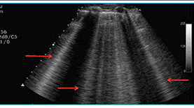
According to radiologist Dr. Robert Bard, indication of the presence of Covid-19 will show in the form of B-LINES (or the 2 vertical rays). "B-Lines specifically mean that the lung tissue adjacent to the coating of the lung is abnormal- and that's what generates these lines. Normal lung tissue should never have B-lines. If you don't have B-Lines, you don't have Covid in the lungs..."

ULTRASOUND ACCESS TO THE "NEW WORLD" OF MEDICINE
The evolution of the portable ultrasound is now being viewed by many as a diagnostic game-changer for patient care in the field. From an interview with professional technical instructor Michael Thury of Terason Ultrasound, he states "the industry just exploded from 30 years ago with the ability and the confidence that ultrasound can give you. The technology enables the physician to really do a much better job diagnosing the patient. As a clinical trainer with clients worldwide, the boom of telemedicine adds to the success of teaching clinicians how to get the most out of remote ultrasound, but also to have fast and easy access to the physician to read and diagnose the patient from any location!"

...............................................................................................................................................................

Seeing your doctor (during the coronavirus pandemic) can be more systematic these days. Just come right to an exam room, get the ultrasound examination, go home, and then review those results with your physician via Video chat.  This third imaging option helps augment radiologic testing for Coronavirus effects in the chest area.

I also see the "bedside ultrasound" in the ICU carry huge benefits use. Being able to bring in a portable unit to a patient's room has a major safety element. Ultrasound can see fluid levels and in densities very nicely and with a small amount of training, you are able to use ultrasound to distinguish the density of the fluid in the areas where there is most fluid. This kind of imaging innovation really helped to support the understanding of A.R.D.S. (Acute Respiratory Distress Syndrome) in association with SIRS (Systemic Inflammatory Response Syndrome) where the lungs are majorly affected by these types of illnesses, filling them with fluid.

For regular monitoring of the lungs, ultrasound is a safe (non radiation) bet.  To identify symptoms in the lungs where the fluid can fill up is priceless data as part of early detection. Failure to breathe happens next, where ventilators come in to increase the oxygen level and be able to push through that fluid all caused by this simple little virus that just goes in and wreaks havoc on a small number of patients.

Percentage wise (not small total number), over 60,000 patients had that high reaction and it loops right back to "how do we keep an eye on that"?  Let's say you had grown a virus and then you're discharged. The lung reaction afterwards can be weeks or months until all the inflammation has gone. Some people less, some people more. And if chest X rays don't really see that type of level of fluid and and infiltration of the lungs with water and if cat scans or a lot of radiation, then these outpatient ultrasound sites could provide a literal progression from the discharge date all the way out until it looks like a normal lung again.

To me, that's a sensible protocol for early detection.


..............................................................................................................................................................

ABOUT THE AUTHOR


STEPHEN CHAGARES, MD, FACS - CANCER SURGEON
Dr. Chagares is a board certified general, laparoscopic, robotic and breast surgeon in Monmouth County, NJ.  He obtained specialty training in breast surgery at Memorial Sloan Kettering Cancer Center. In addition to breast surgery, Dr. Chagares regularly performs hernia repairs of all types, including open, laparoscopic and robotic repairs. He was the first surgeon in his region to perform a robotic hernia repair and remains on the cutting edge of all advanced surgical techniques.  He routinely performs multiple other abdominal procedures, including laparoscopic gallbladder removal.  Dr. Chagares has received numerous awards for excellence and academic teaching, Top Doctors Awards and Patients’ Choice Awards. His philosophy is to provide quality care with a personal touch. He respects the art of surgery and feels honored to treat patients every day. visit his website- drchagares.com


EDITOR / CO-PUBLISHER


ROBERT L. BARD, MD, PC, DABR, FASLMS  - RADIOLOGIST
Dr. Bard is recognized for his specialized work in advanced cancer diagnostic imaging. He co-founded the 9/11 CancerScan program to bring additional diagnostic support to all first responders from Ground Zero. His main practice in midtown, NYC (Bard Diagnostic Imaging- www.CancerScan.com) uses the latest in digital imaging technology and has been also used to help guide biopsies and in many cases, even replicate much of the same reports of a clinical invasive biopsy. Imaging solutions such as high-powered sonograms, Power Doppler Histogram, sonofluoroscopy, 3D/4D image reconstruction and the Power Doppler Histogram  are safe, noninvasive, and do not use ionizing radiation. It is used as a complement to find anomalies and help diagnose the causes of pain, swelling and infection in the body’s internal organs while allowing the diagnostician the ability to zoom and ‘travel’ deep into the body for maximum exploration.


Special thanks to: Alice Chiang and Michael Thury of Terason Ultrasound (https://www.terason.com/) and LonShine Technologies Inc.

Disclaimer & Copyright Notice: The materials provided on this website are copyrighted and the intellectual property of the publishers/producers (The NY Cancer Resource Alliance/IntermediaWorx inc. and Bard Diagnostic Research & Educational Programs). It is provided publicly strictly for informational purposes within non-commercial use and not for purposes of resale, distribution, public display or performance. Unless otherwise indicated on this web based page, sharing, re-posting, re-publishing of this work is strictly prohibited without due permission from the publishers.  Also, certain content may be licensed from third-parties. The licenses for some of this Content may contain additional terms. When such Content licenses contain additional terms, we will make these terms available to you on those pages (which his incorporated herein by reference).The publishers/producers of this site and its contents such as videos, graphics, text, and other materials published are not intended to be a substitute for professional medical advice, diagnosis, or treatment. For any questions you may have regarding a medical condition, please always seek the advice of your physician or a qualified health provider. Do not postpone or disregard any professional medical advice over something you may have seen or read on this website. If you think you may have a medical emergency, call your doctor or 9-1-1 immediately.  This website does not support, endorse or recommend any specific products, tests, physicians, procedures, treatment opinions or other information that may be mentioned on this site. Referencing any content or information seen or published in this website or shared by other visitors of this website is solely at your own risk. The publishers/producers of this Internet web site reserves the right, at its sole discretion, to modify, disable access to, or discontinue, temporarily or permanently, all or any part of this Internet web site or any information contained thereon without liability or notice to you.


Thursday, May 7, 2020

Covid-19 Effects on the Lungs + Chest Ultrasound

Written and presented by: Dr. Stephen Chagares
Edited by: Dr. Robert L. Bard & Lennard Gettz (of NYCRA HealthScan News)

 The Coronavirus has been shown to be very lethal to a subset of patients. What happens is that the virus is ingested into the mouth or nose, going down into the back of the throat (called the hypopharynx and the larynx) and then is breathed into the lungs themselves. The lungs are made of millions of little air SACS called alveoli, and the virus gets into these little layer sacks and adheres to the inner lining. That lining is what allows the interface between the blood and the air and the CO2 from the blood is given out. The oxygen from the air is given into the blood. That's how we breathe in oxygen and blow out CO2.

When the virus gets in there and coats that surface, everyone creates some kind of reaction. One is an antibiotic body reaction- which is an immune response. The other thing that happens is a physical inflammatory response where the immune system automatically goes into defense mode and wants to go in and kill the virus. It senses it's there and sends in "defender" cells, and to most people, it can send the proper amount of little soldiers to do the job. But in a small percentage of patients, the body's internal reaction causes too much inflammation causing a large body of fluid to fill up in the alveoli (air sacs) in the lungs. This stops the ability for oxygen to fuse through those air sacks to get oxygen into the entire body, include vital organs like the heart, the brain, the liver etc.  Without oxygen, this could result serious problems like heart and liver failure (and other such symptoms).

To visually identify this problem, existing technologies have to be tuned to track and monitor cardio-pulmonary performance. Chest x-rays or CT/catscans are available and are widely used now- but carry a number of key issues when it comes to regular chest monitoring. Often normal chest X rays are going to miss this because radiation itself will go through the lung tissue and the fluid and it may not show anything too specific. And then a cat scan may show something totally different where you can see how both lungs are all inflamed and have fluid in them.

The newest thought is the "LUNGSCAN" paradigm through the  use of ultrasound technology- as published by Dr. Robert Bard and a number of European experts in this field. The system of checking the lungs for covid-related issues is applied in tandem with (and not instead of) replacing other radiologic testing.  The concept of INTEGRATIVE solutions is about making as many solutions available to the public as you can to support patient treatment. So although chest X rays and cat scans are often used, there's a lot of radiation to them- and that's a major discrepancy to regular testing.

THE COMPLEX PROCESS OF CT IMAGING
If you're in the ICU, taking a "field trip" down to a radiology center is no fun at all.  It's probably the most dangerous part of a patient's day. Just getting switched from everything on the wall to everything portable and then getting them down and transferring them first from their bed to the stretcher, and then another stretcher into the cat scan table- can be quite harrowing. And then everyone has to get out and leave the patient there. And all the tubes still have to work. And as they come through to make sure nothing pops off and then they have to get transferred back onto the stretcher, up into the elevator, back into the ICU, back into their bed, and then hook everything back up again. So yes, cat scans work great, but there's a lot to it.

PORTABLE IS "IN"
Though we still have the high-powered center units the size of a washing machine, today's ultrasound technology design has been streamlined to offer PORTABLE versions to respond to its many field applications and demands. An "all-in-one" touch screen with detachable probes can be used at the bedside or in any kind of radiologic facility. It is also used in triage units, battlefields, ocean liners, helivacs/ambulances and even the space station.

You can expect to see "LUNGSCAN STATIONS" open up everywhere for outpatients to get quick lung scans (about a 5-minute experience) in urgent care centers per se.  The accuracy, speed and low prep combination of ultrasound alongside our new TeleMedicine trend makes for a SAFER alternative to imaging.


Ultrasound for Critical Care and Regular Covid Monitoring of Respiratory Pathogens

Adding to the arsenal of diagnostic solutions for the Covid-19 respiratory pathogen, the LUNG (or CHEST) ULTRASOUND is making great strides in triage facilities and bedside monitoring.  A growing community of European health specialists are joining a procedural movement to "replace" the dependency on stethoscopes with portable ultrasound use to check a patient's immediate cardiopulmonary condition. For Covid-positive patients, today's ultrasound excels in getting immediate answers safely without radiation - allowing for screening and repeated monitoring. Such is the case in this test sample.

According to radiologist Dr. Robert Bard, indication of the presence of Covid-19 will show in the form of B-LINES (or the 2 vertical rays). "B-Lines specifically mean that the lung tissue adjacent to the coating of the lung is abnormal- and that's what generates these lines. Normal lung tissue should never have B-lines. If you don't have B-Lines, you don't have Covid in the lungs..."

ULTRASOUND ACCESS TO THE "NEW WORLD" OF MEDICINE
The evolution of the portable ultrasound is now being viewed by many as a diagnostic game-changer for patient care in the field. From an interview with professional technical instructor Michael Thury of Terason Ultrasound, he states "the industry just exploded from 30 years ago with the ability and the confidence that ultrasound can give you. The technology enables the physician to really do a much better job diagnosing the patient. As a clinical trainer with clients worldwide, the boom of telemedicine adds to the success of teaching clinicians how to get the most out of remote ultrasound, but also to have fast and easy access to the physician to read and diagnose the patient from any location!"

...............................................................................................................................................................

Seeing your doctor (during the coronavirus pandemic) can be more systematic these days. Just come right to an exam room, get the ultrasound examination, go home, and then review those results with your physician via Video chat.  This third imaging option helps augment radiologic testing for Coronavirus effects in the chest area.

I also see the "bedside ultrasound" in the ICU carry huge benefits use. Being able to bring in a portable unit to a patient's room has a major safety element. Ultrasound can see fluid levels and in densities very nicely and with a small amount of training, you are able to use ultrasound to distinguish the density of the fluid in the areas where there is most fluid. This kind of imaging innovation really helped to support the understanding of A.R.D.S. (Acute Respiratory Distress Syndrome) in association with SIRS (Systemic Inflammatory Response Syndrome) where the lungs are majorly affected by these types of illnesses, filling them with fluid.

For regular monitoring of the lungs, ultrasound is a safe (non radiation) bet.  To identify symptoms in the lungs where the fluid can fill up is priceless data as part of early detection. Failure to breathe happens next, where ventilators come in to increase the oxygen level and be able to push through that fluid all caused by this simple little virus that just goes in and wreaks havoc on a small number of patients.

Percentage wise (not small total number), over 60,000 patients had that high reaction and it loops right back to "how do we keep an eye on that"?  Let's say you had grown a virus and then you're discharged. The lung reaction afterwards can be weeks or months until all the inflammation has gone. Some people less, some people more. And if chest X rays don't really see that type of level of fluid and and infiltration of the lungs with water and if cat scans or a lot of radiation, then these outpatient ultrasound sites could provide a literal progression from the discharge date all the way out until it looks like a normal lung again.

To me, that's a sensible protocol for early detection.


..............................................................................................................................................................

ABOUT THE AUTHOR


STEPHEN CHAGARES, MD, FACS - CANCER SURGEON
Dr. Chagares is a board certified general, laparoscopic, robotic and breast surgeon in Monmouth County, NJ.  He obtained specialty training in breast surgery at Memorial Sloan Kettering Cancer Center. In addition to breast surgery, Dr. Chagares regularly performs hernia repairs of all types, including open, laparoscopic and robotic repairs. He was the first surgeon in his region to perform a robotic hernia repair and remains on the cutting edge of all advanced surgical techniques.  He routinely performs multiple other abdominal procedures, including laparoscopic gallbladder removal.  Dr. Chagares has received numerous awards for excellence and academic teaching, Top Doctors Awards and Patients’ Choice Awards. His philosophy is to provide quality care with a personal touch. He respects the art of surgery and feels honored to treat patients every day. visit his website- drchagares.com


EDITOR / CO-PUBLISHER


ROBERT L. BARD, MD, PC, DABR, FASLMS  - RADIOLOGIST
Dr. Bard is recognized for his specialized work in advanced cancer diagnostic imaging. He co-founded the 9/11 CancerScan program to bring additional diagnostic support to all first responders from Ground Zero. His main practice in midtown, NYC (Bard Diagnostic Imaging- www.CancerScan.com) uses the latest in digital imaging technology and has been also used to help guide biopsies and in many cases, even replicate much of the same reports of a clinical invasive biopsy. Imaging solutions such as high-powered sonograms, Power Doppler Histogram, sonofluoroscopy, 3D/4D image reconstruction and the Power Doppler Histogram  are safe, noninvasive, and do not use ionizing radiation. It is used as a complement to find anomalies and help diagnose the causes of pain, swelling and infection in the body’s internal organs while allowing the diagnostician the ability to zoom and ‘travel’ deep into the body for maximum exploration.


Special thanks to: Alice Chiang and Michael Thury of Terason Ultrasound (https://www.terason.com/) and LonShine Technologies Inc.

Disclaimer & Copyright Notice: The materials provided on this website are copyrighted and the intellectual property of the publishers/producers (The NY Cancer Resource Alliance/IntermediaWorx inc. and Bard Diagnostic Research & Educational Programs). It is provided publicly strictly for informational purposes within non-commercial use and not for purposes of resale, distribution, public display or performance. Unless otherwise indicated on this web based page, sharing, re-posting, re-publishing of this work is strictly prohibited without due permission from the publishers.  Also, certain content may be licensed from third-parties. The licenses for some of this Content may contain additional terms. When such Content licenses contain additional terms, we will make these terms available to you on those pages (which his incorporated herein by reference).The publishers/producers of this site and its contents such as videos, graphics, text, and other materials published are not intended to be a substitute for professional medical advice, diagnosis, or treatment. For any questions you may have regarding a medical condition, please always seek the advice of your physician or a qualified health provider. Do not postpone or disregard any professional medical advice over something you may have seen or read on this website. If you think you may have a medical emergency, call your doctor or 9-1-1 immediately.  This website does not support, endorse or recommend any specific products, tests, physicians, procedures, treatment opinions or other information that may be mentioned on this site. Referencing any content or information seen or published in this website or shared by other visitors of this website is solely at your own risk. The publishers/producers of this Internet web site reserves the right, at its sole discretion, to modify, disable access to, or discontinue, temporarily or permanently, all or any part of this Internet web site or any information contained thereon without liability or notice to you.

©Copyright Intermedia Worx Inc./Prevention 101. All Rights Reserved.

Thursday, April 2, 2020

EUROPEAN IMAGING COLLEAGUES CONFIRM LUNG ULTRASOUND BENEFITS FOR COVID-19 TRIAGE

April 2, 2020 - Dr. Robert L. Bard, NYC imaging specialist collaborates with an international group of medical leaders including Dr. Danilo Buonsenso (Rome, Italy) who recently published reports about "ultrasound equipment as an effective replacement of the stethoscope.”  Dr. Buonsenso's review presents the valid uses and benefits of Lung Ultrasound in identifying respiratory disorders that may be associated with Covid-19.

Currently, front line physicians in Italy and Spain are reportedly triaging with portable ultrasound units that reduce logistical problems of a chaotic environment and healthcare worker exposure. Since imaging with CT or ultrasound is not diagnostic, determining who needs hospitalization is essential in a pandemic overwhelming medical providers.  Dr. Buonsenso, at the viral epicenter in Rome, uses sonograms to decide who requires a CT scan.

Sonogram taken under rib cage shows liver (grey) with curved diaphragm-
lung border (white) Arrows point to vertical B lines (white) demonstrating
diseased lung tissue.  The more B lines the worse the disease. Healing is
measured by reduction in the number of B lines
The use of CT lung imaging for COVID-19 has been recognized as the diagnostic standard during our current epidemic. Meanwhile, experts find CT to have  disadvantages like radiation exposure. Respiratory distress creates motion artifacts on images that may simulate pulmonary inflammation. Also, viral pneumonia is not diagnostically distinguishable from other viral inflammations in the lung so the argument for a screening modality is useful to separate the critically ill from those needing outpatient treatment.  This review was stated by Dr. Klaus Lessnau, author of "Atlas of Chest Sonography" (Springer 2003), employs both CT and ultrasound imaging in clinical practice.

Dr. Bard reviews international healthcare and technology updates as part of his continued research in the radiological society. This includes Dr. Buonsenso's national reports about the Covid crisis and the expanded use of lung ultrasound as part of his investigation of children as clinically unaffected carriers.  “The global pandemic demands effective answers toward a cure as well as protection of healthcare workers on duty", says Dr. Bard. "I have the highest regard for the European approach to problem solving both clinically and technologically… applying Dr. Buonsenso’s concept makes perfect sense to me and carries great value in our war against Covid-19.”

Ultrasound probes study the lungs between the ribs to
read the lung surface where most Covid pathology is situated
According to Dr. Bard, Lung ultrasound has been used in emergency rooms since it was introduced to the Mt Sinai Medical School in 2014 and is now used nationwide to diagnose pneumonia (viral or bacterial) in children which spares them unnecessary x-rays since it is so accurate. “It is like an electronic stethoscope since lung disease and heart failure producing pulmonary fluid buildup are diagnosed or confirmed with portable ultrasound units at the bedside. This is considered the best imaging tool to diagnose a collapsed lung in seconds which has proven lifesaving as a time saver for on the spot detection.”

In a recent telehealth conference, Dr. Bard explains how ultrasound probes study the lungs between the ribs to read the lung surface where most Covid pathology is situated.  (Image 1) Portable units have the advantage of containment within a sterile sleeve, preventing accidental viral spread to imaging equipment necessitating full decontamination procedures.  All clinical imaging was correlated with the patient’s oxygen saturation and clinical setting.     The virus has known cardiac toxicity so the same sonogram unit may image the heart for fluid buildup and weakened contraction. This may differentiate heart failure from pulmonary infection in some cases which may have similar clinical presentations.


About Dr. Bard
Dr. Robert Bard currently runs a private imaging center in NYC specializing in advanced 3-D sonography to detect cancer tumors and other health disorders.  He lectures in medical conferences worldwide, runs a cancer awareness program for first responders and is also a publisher of countless educational books and articles about cancer imaging and other health/wellness related materials.

Dr. Bard maintains an active role in supporting the medical community by contributing relevant articles to major health magazines, medical journals and news organizations pertaining to current health concerns. His recent projects include advocating and inserting TELEMEDCINE in the medical community as a safe alternative for patients.  Other projects include an upcoming collaborative textbook series on Covid-19 with a list of top experts in the field.  Dr. Bard is also the president of the AngioFoundation (501c3), as philanthropic organization dedicated to funding and supporting public education about current treatment protocols worldwide. 


About Dr. Buonsenso
Actively practicing as a Pediatrician from Gemelli University Hospital in Italy, Dr. Danilo Buonsenso received his medical degree from the Catholic University of the Sacred Heart in Rome in 2010. He was a resident in The School of Pediatrics in the Catholic University of the Sacred Heart till 2016. From January 2016, he was a Head Director of a social and health developing program in the community of Bureh Town, Sierra Leone which aimed to bring medical and social support to fragile populations. At present, he is a Pediatrician at the Department of Woman and Child Health and Publich Health at the Gemelli University Hospital, Rome, Italy.

Dr. Buonsenso's basic personal skills are in Infectious diseases, Pediatric ER and Pediatric Ultrasonography. He has published almost 50 papers in various journals of Pediatrics and has received various grants such as a Grant as a young collaborator by The Italian Minister of Health for the Bando Ricerca Finalizzata 2018, on the role of NGF on brain function recovery after severe brain injury. (Source)

About Dr. Klaus Lessnau
Dr. Klaus Lessnau specializes in Pulmonology, Sleep Medicine, Insomnia Testing, Sleep Apnea Testing, Pulmonary Function Testing.  He is a highly regarded Pulmonologist and a Sleep Medicine specialist with the 14 Street Medical Arts team in NYC. Dr. Lessnau is board certified in Pulmonology, Bronchology, Sleep Medicine Disorders and is a Critical Care doctor with over 30 years of experience. He is also serving as a Director of Critical Care Medicine at Lennox Hill Hospital.


#    #    #

References
1) Is there a role for lung ultrasound during the COVID‐19 pandemic?  - Danilo Buonsenso https://onlinelibrary.wiley.com/doi/abs/10.1002/jum.15284

2) The Lancet: COVID-19 outbreak: less stethoscope, more ultrasound" https://www.thelancet.com/journals/lanres/article/PIIS2213-2600(20)30120-X/fulltext
Danilo Buonsenso | Davide Pata | Antonio Chiaretti - Published:March 20, 2020

3) Dr. Klaus Lessnau, author of "Atlas of Chest Sonography" (Springer 2003) https://link.springer.com/book/10.1007%2F978-3-662-05278-5


Disclaimer & Copyright Notice: The materials provided on this website are copyrighted and the intellectual property of the publishers/producers (The NY Cancer Resource Alliance/IntermediaWorx inc. and Bard Diagnostic Research & Educational Programs). It is provided publicly strictly for informational purposes within non-commercial use and not for purposes of resale, distribution, public display or performance. Unless otherwise indicated on this web based page, sharing, re-posting, re-publishing of this work is strictly prohibited without due permission from the publishers.  Also, certain content may be licensed from third-parties. The licenses for some of this Content may contain additional terms. When such Content licenses contain additional terms, we will make these terms available to you on those pages (which his incorporated herein by reference).The publishers/producers of this site and its contents such as videos, graphics, text, and other materials published are not intended to be a substitute for professional medical advice, diagnosis, or treatment. For any questions you may have regarding a medical condition, please always seek the advice of your physician or a qualified health provider. Do not postpone or disregard any professional medical advice over something you may have seen or read on this website. If you think you may have a medical emergency, call your doctor or 9-1-1 immediately.  This website does not support, endorse or recommend any specific products, tests, physicians, procedures, treatment opinions or other information that may be mentioned on this site. Referencing any content or information seen or published in this website or shared by other visitors of this website is solely at your own risk. The publishers/producers of this Internet web site reserves the right, at its sole discretion, to modify, disable access to, or discontinue, temporarily or permanently, all or any part of this Internet web site or any information contained thereon without liability or notice to you.

©Copyright Intermedia Worx Inc./Prevention 101. All Rights Reserved.

Tuesday, March 31, 2020

PROTON THERAPY: CANCER TREATMENT TECHNOLOGY IN REVIEW

By: Dr. Robert L. Bard & Grace Davi
Edited by: Lennard M. Gettz / NY Cancer Resource Alliance

Medical researchers and developers have historically pursued many similar considerations in the path to improving cancer treatment solutions—much more than simply "killing cancer tumors."  The highest priority is typically given toward patient safety and well-being during and after treatment due to the use of highly powerful foreign elements like radiation and chemicals with heavy toxicity levels.

Such is the case with conventional X-ray (photon) treatments like intensity-modulated radiation therapy (IMRT), which has proven to be successful in killing targeted tumors but also can damage nearby tissue, thus causing injury to the patient.  IMRT applies high doses of irradiation in order to penetrate the body and reach the depths of the targeted tumor. This powerful beam of energy exposes all tissues along its entire path to radiation, including the normal tissues before the tumor and the normal tissues past the tumor.

 An upgrade from using x-rays in radiation therapy came with the delivery of charged (proton) particle beams (originated by Dr. Robert R. Wilson, 1946) to irradiate cancer. This dose is deposited within a controlled range of depth, affecting specific coordinates in the body so most of the dose is delivered to the actual tumor and little or no radiation is delivered to tissues beyond the tumor (called the Bragg peak). This technique, therefore, maximizes the chances of curing patients without cause debilitating side effects, as proton research shows promising results in reducing the damage to healthy tissues and better preserving patient quality of life.

The National Association for Proton Therapy (NAPT) reports that both standard radiation therapy and proton therapy to work on the same principle of damaging cellular DNA of tumor, with the major difference that proton therapy deposits the majority of the radiation dose directly into the tumor and travels no further through the body.  According to NAPT spokesperson Jennifer Maggiore, “The FDA approved this technology over 30 years ago, so it's not necessarily new, but recent advancements have made it more accessible in hospitals, and versions are also developed for single-room systems.” There are large “big scale” installations with a cyclotron that feeds three to four gantries. This takes up a big footprint of space and a major investment of time and money, which has led to the increase in smaller, single-room centers in recent years.

IMPROVING TRENDS IN CANCER TREATMENT
It is commonly observed that surgeons are increasingly using minimally invasive procedures.  Whether it's robotic or video assisted surgeries, we can identify the pattern of new treatment protocols to result in higher quality of life and a reduction in toxicity. In doing so, it allows us, in some cases, to actually improve survival through those same methods of reducing toxicities for patients.

According to Dr. Charles B. Simone II, Chief Medical Officer of the New York Proton Center, “We’re going to see more and more customized treatment; it's not a one size fits all approach to cancer. We are going to have individualized ways to deliver radiation therapy, individualized drugs or immune agents—and then, potentially more synergy between modalities such as radiation with systemic therapies.”

The concept of the pencil beam scanning or IMPT (intensity-modulated proton therapy) has grown widely accepted as the ‘new future’ in radiation therapy.  Originally recognized to treat brain tumors, proton therapy has since found global success in treating prostate, breast, liver, lung, head and neck, and other cancers.

In the recent past, proton therapy has continued to advance in its design and performance. Over the past two decades, the number of academically affiliated proton therapy centers in the United States has grown from zero to 31.  Over the past six years, newer centers have come onboard with pencil beam scanning proton therapy that has enabled IMPT. This new generation of proton therapy allows the radiation to be focused and deposited directly at the tumor, while avoiding normal tissues to an even greater extent than the first generation of proton therapy. Another unique advantage of the pencil beam scanning includes its ability to better sculpt the beam or dose.  To match the beam into the shape of the tumor (which is usually not a perfectly square, circle or rectangle shape) allows the deposit of more radiation into the tumor, as it travels into the patient, with even less radiation deposited in the normal tissues in front of and also after the tumor.

According to Dr. Simone, another recent advancement in proton therapy is the ability for physicians to apply volumetric imaging—or the ability to conduct low dose CT scans daily and immediately (in 3D) before treatment—to the targeted area. Volumetric imaging allows radiation oncologists to directly visualize the tumor, or the area that needs the treatment, without having to rely on bony anatomy as a surrogate, as most proton treatment installations do. Most proton facilities still use X-ray or KV images, rather than a cone beam CT image, limiting the ability to have millimeter precision.

Unlike devices such as the CyberKnife system with a regular linear accelerator that essentially plugs into the wall and generates its own radiation, proton therapy requires its own source of energy to generate the proton therapy. The most common model used by proton centers to generate protons, including at the New York Proton Center, is through a cyclotron—a 10-foot-wide machine that accelerates particles about two-thirds of the speed of light to generate protons. From there, the radiation gets siphoned out of the cyclotron through a beam line that's just a few inches wide, and goes into each of the clinical treatment rooms.

Proton therapy has been shown to reduce the risk of secondary cancers in patients, while decreasing the chance of any long-term complications from the treatment. For some cancers, including for most pediatric cancers, it has grown to be called the de facto standard of care, while for other cancers clinical trials are being conducted to determine it as the preferred treatment for specific patient populations.

FROM THE PATIENTS’ SIDE
After the patient’s radiation oncologist determines that they are qualified for proton therapy, patients would come in for a single preparation appointment, what's called a simulation or radiation mapping appointment.  This is generally done with an image (like a CT scan, a PET scan or an MRI), where the physician will work with a radiation physicist, as well as treatment planning dosimetrist, to map out the tumor in three or four dimensions.  This helps identify how to deliver radiation to that tumor while avoiding irradiation the normal tissues.

“There are several factors that help us determine the right form of treatment: the type of cancer, the tumor location and other patient characteristics. The length of the treatment varies depending on the case,” explained Dr. Simone. “Some patients will go through stereotactic proton therapy, which is generally between one and five day, and others will experience a more conventional treatment that's every day, Monday through Friday, for several weeks. While most treatments with proton therapy are the same number of days as with traditional x-ray therapy, because of the ability for protons to limit side effects, in some cases proton therapy can be administered to patients in high doses per day, leading to shorter treatment times, decreased cost, more patient convenience, and in some cancers better chances of cure.”


THE NEW YORK PROTON CENTER
July 2019 marked the opening of the 140,000 square ft. state of the art proton treatment facility on East 126th Street.  Managed by ProHealth medical group, the New York Proton Center was established under a joint partnership between Memorial Sloan Kettering Cancer Center, Mt. Sinai Health and Montefiore Health System. The New York Proton Center is projected to treat approximately 1,400 patients annually, receiving patients from its consortium partner institutions and from patients throughout the New York metro area and beyond who are looking for the most effective radiation care possible. The center will be one of the few worldwide that is equipped with the newest and most effective proton therapy technology, provided by globally renowned Varian Medical Systems, the worldwide leader in developing multidisciplinary, integrated cancer solutions.


ABOUT DR. SIMONE
Dr. Charles B. Simone, II is the Chief Medical Officer of the New York Proton Center. He is an internationally recognized expert in the use of proton therapy to treat thoracic malignancies and for reirradiation, and in the development of clinical trial strategies and innovative research in thoracic radiation oncology and stereotactic body radiation therapy. He is a National Institutes of Health, National Science Foundation, and Department of Defense funded investigator who performs clinical and translational research investigating the benefits of proton therapy as part of multi-modality therapy for thoracic malignancies. After years of dedication and service to the American College of Radiation Oncologists, Dr. Simone has been named a Fellow of ACRO, recognizing his highly valued contributions to the field. He has published over 340 scientific articles and chapters, given over 210 scientific lectures to national and international audiences, and is the national Principal Investigator or Co-Chair of 7 NIH-funded cooperative group trials (see complete bio- link https://www.nyproton.com/charles-simone/)


CONTRIBUTORS /  EDITORIAL TEAM

ROBERT L. BARD, MD, PC, DABR, FASLMS - Advanced Imaging & Diagnostic Specialist
Having paved the way for the study of various cancers both clinically and academically, Dr. Robert Bard co-founded the 9/11 CancerScan program to bring additional diagnostic support to all first responders from Ground Zero. His main practice in midtown, NYC (Bard Diagnostic Imaging- www.CancerScan.com) uses the latest in digital Imaging technology has been also used to help guide biopsies and in many cases, even replicate much of the same reports of a clinical invasive biopsy. Imaging solutions such as high-powered Sonograms, Spectral Doppler, sonofluoroscopy, 3D/4D Image Reconstruction and the Spectral Doppler are safe, noninvasive, and does not use ionizing radiation. It is used as a complement to find anomalies and help diagnose the causes of pain, swelling and infection in the body’s internal organs while allowing the diagnostician the ability to zoom and ‘travel’ deep into the body for maximum exploration.

GRACE DAVI, Public Health Research Consultant at The RightWriters Group 
Grace dedicated her life's work to intense reporting and data analyses of Cancer-related environmental issues. In addition to content work Grace is also a public advocate for health and safety projects in professional areas and support programs for Infection Prevention branches of health care.  Grace launched her career as a researcher/reporter by pioneering collaborative lab projects in the New York waterways by providing public awareness about contaminants and leaching into county and state aquifer. She combined this experience with   4+ years working with oncologists and cancer immunologists as an editor in medical education. Today, Grace is one of the editors and co-publishers of health related publcations, websites, newsletters and journals including prevention101.org and ImmunologyFirst.org


Special Thanks
The NY Cancer Resource Alliance writing team and AngioMedical Publishing wishes to express its deepest and most heartfelt thanks to Dr. Charles B. Simone II for his kind generosity in sharing his vast knowledge about the science and technology of Proton Therapy.  Special added appreciation also belongs to
 the staff at The NY Proton Center including Patrick Curry and Miriam Mond for their support, and also to Nathaniel Goehring of Berlin Rosen Public Relations and Jane Fort and Jennifer Maggiore of the National Association of Proton Therapy (NAPT) - without whose coordinated efforts this project would not have been possible. 


References:
1) https://www.modernhealthcare.com/providers/proton-center-set-open-new-york
2) https://www.itnonline.com/article/trends-proton-therapy-%E2%80%94-faster-therapy-delivery-single-room-installs
3) https://www.manhattantimesnews.com/proton-powerpoder-de-protones/
4) https://www.mevion.com/newsroom/press-releases/mevion-s250-becomes-first-proton-therapy-system-approved-treat-cancer
5) Video: https://www.youtube.com/watch?time_continue=7&v=MS590Xtq9M4&feature=emb_title


Disclaimer & Copyright Notice: The materials provided on this website are copyrighted and the intellectual property of the publishers/producers (The NY Cancer Resource Alliance/IntermediaWorx inc. and Bard Diagnostic Research & Educational Programs). It is provided publicly strictly for informational purposes within non-commercial use and not for purposes of resale, distribution, public display or performance. Unless otherwise indicated on this web based page, sharing, re-posting, re-publishing of this work is strictly prohibited without due permission from the publishers.  Also, certain content may be licensed from third-parties. The licenses for some of this Content may contain additional terms. When such Content licenses contain additional terms, we will make these terms available to you on those pages (which his incorporated herein by reference).The publishers/producers of this site and its contents such as videos, graphics, text, and other materials published are not intended to be a substitute for professional medical advice, diagnosis, or treatment. For any questions you may have regarding a medical condition, please always seek the advice of your physician or a qualified health provider. Do not postpone or disregard any professional medical advice over something you may have seen or read on this website. If you think you may have a medical emergency, call your doctor or 9-1-1 immediately.  This website does not support, endorse or recommend any specific products, tests, physicians, procedures, treatment opinions or other information that may be mentioned on this site. Referencing any content or information seen or published in this website or shared by other visitors of this website is solely at your own risk. The publishers/producers of this Internet web site reserves the right, at its sole discretion, to modify, disable access to, or discontinue, temporarily or permanently, all or any part of this Internet web site or any information contained thereon without liability or notice to you.

Monday, March 30, 2020

TRIAGE OF COVID-19 BY LUNG ULTRASOUND

 by Robert  Bard MD, FASL  with contributors: Dr. K. Lessnau, New York with Dr. D. Buonsenso, Rome

The use of CT lung imaging for COVID-19 has been the diagnostic standard for the last few months of the current epidemic.  CT has the disadvantage of logistics (staff, patients, transport) and radiation exposure. Respiratory distress creates motion artifacts on images that may simulate pulmonary inflammation. Since the disease mutates often, frequent imaging may be required. Viral pneumonia is not diagnostically distinguishable from other viral inflammations in the lung so the argument for a screening modality is useful to separate the critically ill from those needing outpatient treatment.  Dr. Klaus Lessnau, author of CHEST ULTRASOUND (Springer 2003), employs both CT and ultrasound imaging in clinical practice.


Ultrasound probes have the ability to screen the lungs for respiratory
issues and is a useful TRIAGE tool- however no radiological device
has been able to identify pulmonary viruses directly
Front line physicians in Italy and Spain are triaging with portable bedside ultrasound units that reduce logistical problems of a chaotic environment and healthcare worker exposure. Since imaging with CT or ultrasound is not diagnostic, determining who needs hospitalization is essential in a pandemic that is overwhelming medical providers and hospital resources.  Dr. Buonsenso, on the front lines in Rome, uses sonograms to decide who gets a CT scan. This is key since deep cleaning a CT room after a suspected patient shuts the room down for up to 2 hours for decontamination.

Lung ultrasound has been used in emergency rooms since it was introduced to the Mt Sinai Med School (New York) Emergency Department in 2014 and is now used nationwide to diagnose pneumonia (viral or bacterial) in children which spares them unnecessary x-rays since it is so caccurate. It is like an electronic stethoscope since lung disease and heart failure producing pulmonary fluid buildup are diagnosed or confirmed with portable ultrasound units at the bedside. This is considered the best imaging tool to diagnose a collapsed lung in seconds which has proven lifesaving as a time saver for on the spot detection. While it was assumed that children are carriers and not clinically affected, Dr. Buonsenso is actively investigating this population and there are findings that are concerning with the expanded use of lung ultrasound in this understudied group.

Disease of the lung from fluid overload-infection, 
heart failure-produces vertical white lines (B-Lines)
Ultrasound probes study the lungs via the ribs showing the lung surface (where most Covid pathology is situated) and adjacent lung tissue. The abdominal scan with the curved transducer has a larger field of view and affords a rapid assessment of B lines (Fig 1), pleural effusion and frank pneumonia (Fig 2).  Portable units have the advantage of containment within a sterile sleeve preventing accidental viral spread to imaging equipment necessitating full decontamination procedures.  Some infected European physicians are monitoring their disease at home via the B line count-few B lines suggest low grade inflamed lung tissue-increasing B line count calls for more aggressive treatment. All clinical imaging was correlated with the patient’s oxygen saturation and clinical setting.  The virus has potential cardiac toxicity so the same sonogram unit may image the heart for fluid buildup and weakened contraction. This may differentiate heart failure from pulmonary infection in some cases which may have similar clinical presentations.

REFERENCE
Bard R: 3D Imaging of Pulmonary Edema in Proceedings of 2020 Annual  American Institute of Ultrasound in Medicine ;Supplement to Journal of Ultrasound in Medicine  July 2020 (in press)

.............................................................................................................................................................

CONTRIBUTING WRITER

ROBERT L. BARD, MD, PC, DABR, FASLMS - Advanced Imaging & Diagnostic Specialist
Having paved the way for the study of various cancers both clinically and academically, Dr. Robert Bard co-founded the 9/11 CancerScan program to bring additional diagnostic support to all first responders from Ground Zero. His main practice in midtown, NYC (Bard Diagnostic Imaging- www.CancerScan.com) uses the latest in digital Imaging technology has been also used to help guide biopsies and in many cases, even replicate much of the same reports of a clinical invasive biopsy. Imaging solutions such as high-powered Sonograms, Spectral Doppler, sonofluoroscopy, 3D/4D Image Reconstruction and the Spectral Doppler are safe, noninvasive, and does not use ionizing radiation. It is used as a complement to find anomalies and help diagnose the causes of pain, swelling and infection in the body’s internal organs while allowing the diagnostician the ability to zoom and ‘travel’ deep into the body for maximum exploration.


Disclaimer & Copyright Notice: The materials provided on this website are copyrighted and the intellectual property of the publishers/producers (The NY Cancer Resource Alliance/IntermediaWorx inc. and Bard Diagnostic Research & Educational Programs). It is provided publicly strictly for informational purposes within non-commercial use and not for purposes of resale, distribution, public display or performance. Unless otherwise indicated on this web based page, sharing, re-posting, re-publishing of this work is strictly prohibited without due permission from the publishers.  Also, certain content may be licensed from third-parties. The licenses for some of this Content may contain additional terms. When such Content licenses contain additional terms, we will make these terms available to you on those pages (which his incorporated herein by reference).The publishers/producers of this site and its contents such as videos, graphics, text, and other materials published are not intended to be a substitute for professional medical advice, diagnosis, or treatment. For any questions you may have regarding a medical condition, please always seek the advice of your physician or a qualified health provider. Do not postpone or disregard any professional medical advice over something you may have seen or read on this website. If you think you may have a medical emergency, call your doctor or 9-1-1 immediately.  This website does not support, endorse or recommend any specific products, tests, physicians, procedures, treatment opinions or other information that may be mentioned on this site. Referencing any content or information seen or published in this website or shared by other visitors of this website is solely at your own risk. The publishers/producers of this Internet web site reserves the right, at its sole discretion, to modify, disable access to, or discontinue, temporarily or permanently, all or any part of this Internet web site or any information contained thereon without liability or notice to you.