Wednesday, July 24, 2019

9/11 Asthma Cases & the Firefighters' Cough Continues to Plague First Responders

© Copyright 2019 - The Biofoundation for AngioGenesis R&D / IntermediaWorx inc. All Rights Reserved. Published for the NY Cancer Resource Alliance (NYCRA), AwarenessforaCure.org and HealthScanNYC.org


FOREWORD by: Dr. Jesse A. Stoff
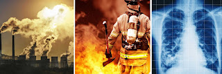
Recently in the news, they're talking a lot about asbestos and asbestos-related cancers due to 9/11 exposure because when the Twin Towers collapsed, all the asbestos that was in there for insulation was aerosolized. And when you breathe that stuff in, in small particles that have been micronized from the explosion and compression phenomena, when those particles get lodged in the lungs, the body doesn't have a good way to excrete it.   Because lung tissue (unlike liver tissue for example) heals by scarring and not regeneration, when the lungs are exposed to chronic irritants that the body can't get rid of, chronic inflammation and irritation ultimately leads to the death of lung cells called pneumocytes.  That area of damage causes bronchiectasis and scar tissue formation which can lead to COPD and the diseases associated with that including cardiovascular problems and death.  [1] (source: Huntington Patch)


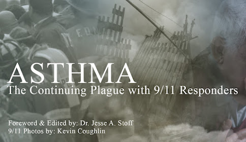
ASTHMA: A MAJOR PREVALENCE WITH FIRST 9/11 RESPONDERS
Fact: no two individuals are ever the same especially when it comes to the physiological effects of envrionmental health hazards- such as those from a disaster zone like Ground Zero. We have all seen countless cases of health issues appearing for the first time 10-15 years after 2001, and the same includes respiratory disorders like ASTHMA.

Where logic may dictate that  the giant plume of noxious dust should equate to a widepsread case of pulmonary issues within moments of contact, physicians have observed a variety of effects depending on body types (reflecting genetic makeup) or possibly a unique tolerance level that may actually resist or even 'hide' any symptoms until well past a decade from the exposure.  Others may even continue to show zero evidence of negative effects at all (or for now).

"THE TELLTALE COUGH"- EXPLAINED
According to Dr. Paul Schulster, (pulmonologist from Oceanside, NY) the COUGH can say a lot, but often misleads the patient as a "nothing" or a "simple little cough".  For firefighters, it is usually a telltale sign of various possible issues. The first syndrome often comes from a post-nasal drip. The second most common cause is from irritation, inflammation and bronchiospasm. Third is Gastroesophageal Reflux Disease. My 9/11-related patients that have GERD starts with that warning cough while others' coughs can trigger the asthma.  Finally, Irritative Cough Syndrome can also happen where one cough leads to another cough, irritating the airway, exacerbating another cough - and then another.

Having a cough here or a wheeze there is not enough for most first responders to raise the flag of alarm. Seasoned specialists like Dr. Schulster recognizes that unique and unusual symptoms or maladies do not reach the patient's consciousness for quite some time.  Ignoring or not paying more attention to these "little" anomalies tend to often be the norm.  These coughs may progressively grow worse over the years and then one day they begin to wheeze a little more than usual and wind up with advancing shortness of breath.  Once this becomes significant and finally enters their consciousness, only then will the thought of seeking medical help actually come to mind.


DIAGNOSTIC OPTIONS
Oftentimes, an exam from the pulmonologist starts with the CAT scans of the chest. The firefighters are being tracked for pulmonary nodules. They're referred to as sub-centimeter nodules, which are so small that you can't read it. "You don't really see them on a plain X-ray, chest X-rays, PA and lateral. A lot of these first responders already come to me with CAT scans from the past and have been followed by World Trade Center program and the FDNY doctors that are also pulmonary doctors"- states Dr. Schulster.

In a pulmonologist's tool kit exists certain standard pulmonary function examss- including the SPIROMETRY [2].  This test estimates the narrowing of your bronchial tubes by checking how much air you can exhale after a deep breath and how fast you can breathe out [5].   This allows us to  see the best way of determining the lung function in numbers, more or less, is a complete pulmonary function test.  Next is the METHACHOLINE CHALLENGE [3] - also known as an asthma trigger that, when inhaled, will cause mild constriction of your airways.  If you react to the methacholine, you likely have asthma. This test may be used even if your initial lung function test is normal. [5]   Another test used is THE COLD AIR CHALLENGE [4]. The  patients generally come with having had those in the past and most are positive for asthma. In the asthmatics. 

Inevitably, multiple poisons inhaled in 'the pile' trigger disorders that are obtained on a longterm basis. The isocyanates and the aldehyde may trigger the asthma, "but I'm not certain if we really know the specific cause of their 9/11 based asthma. There's a long list of toxins that irritate and inflame. The probable causes of Asthma are either chronic of acute inflammation. As they breathed in the 9/11 dust, they breathed in 30 of those toxins, causing inflammation in the airways which then led to chronic reactions."

The sub-centimeter nodules seems to be frequent with 9/11 responders. The good news is that most of them turn out to be benign.  One follows these nodules for a couple of years with images and CAT scans because they're often too small to really see on plain chest X-rays. And if they remain the same size, they get smaller over a few years, then they're considered benign. And then that's how we deal with it.

Concluding Dr. Schulster's interview, we found that identifying a chronic respiratory disorder like Asthma can be quite involved that there are various diagnostic solutions and treatment options available depending on its classification or severity. Especially in the case of a first responder's long-term exposure to toxic fumes, recognizing the source(s) of contamination can greatly help the physician establish the proper treatment strategy for the patient.


EXTRA: ASTHMA TREATMENT OPTIONS  
source: https://www.mayoclinic.org/diseases-conditions/asthma/diagnosis-treatment/drc-20369660
Prevention and long-term control are key in stopping asthma attacks before they start. Treatment usually involves learning to recognize your triggers, taking steps to avoid them and tracking your breathing to make sure your daily asthma medications are keeping symptoms under control. In case of an asthma flare-up, you may need to use a quick-relief inhaler, such as albuterol.

Medications
The right medications for you depend on a number of things — your age, symptoms, asthma triggers and what works best to keep your asthma under control. Preventive, long-term control medications reduce the inflammation in your airways that leads to symptoms. Quick-relief inhalers (bronchodilators) quickly open swollen airways that are limiting breathing. In some cases, allergy medications are necessary. Long-term asthma control medications, generally taken daily, are the cornerstone of asthma treatment. These medications keep asthma under control on a day-to-day basis and make it less likely you'll have an asthma attack.  See complete list of TREATMENT options and full descriptions @ MAYO CLINIC's website:
https://www.mayoclinic.org/diseases-conditions/asthma/diagnosis-treatment/drc-20369660

..................................................................................................................................................................

STAFF EDITOR 
JESSE STOFF, MD, HMD, FAAFP is a highly-credentialed medical expert studying all medical remedies in pursuit of resolving the most challenging health issues of our time. In many circles, he is recognized for his 35+ years of dedicated work in immunology and advanced clinical research in modern CANCER treatments. He has spoken worldwide in some of the most sought-after medical conferences about his experiences and analyses on the study of human disease. His integrative practice (INTEGRATIVE MEDICINE OF NY, Westbury, NY) has been continually providing all patients with the many comprehensive clinical options and modalities available- including "ONCO-IMMUNOLOGY", the science of battling cancer cells and reversing pre-cancerous conditions through a complete prevention program that has earned him great success in this field.  For more information, visit: www.Dr.JesseStoff.com


CONTRIBUTING 9/11 PHOTOGRAPHER
KEVIN P. COUGHLIN is a Pulitzer Prize-sharing photojournalist, writer, director of photography, pilot, and aerial cinematographer. He is the current executive photographer to New York Governor Andrew M. Cuomo. His photographs at Ground Zero following the September 11, 2001 attacks on the World Trade Center and while covering funerals and memorial services of fallen fire fighters, police officers, and emergency personnel killed as a result of the attacks are included in the 2002 Pulitzer Prize awarded to The New York Times for Public Service. In addition to The New York Times, his photographs have appeared in the New York Post, New York Daily News, Newsday, The Philadelphia Inquirer,  https://www.kevincoughlinphotography.com/


PROFESSIONAL INTERVIEWED IN THIS ARTICLE
PAUL L. SCHULSTER, MD PC is a practicing Pulmonary Disease Specialist in Oceanside, NY. Dr. Schulster graduated from University of Kentucky College of Medicine in 1972 and has been in practice for 47 years. He completed a residency at Queens Hospital Center. Dr. Schulster also specializes in Internal Medicine. Dr. Schulster also practices at South Nassau Community Hospital. One Healthy Way Oceanside NY. His private practice is located at: 442 Waukena Avenue, Oceanside, New York. 11572 |  (516) 599-8234


References:
1)The 9/11 Attacks are Still Going On with Asbestos Based Cancers- by: Jesse Stoffhttps://patch.com/new-york/huntington/9-11-attacks-are-still-going-asbestos-based-cancers
2) Spirometry: https://www.healthline.com/health/spirometry
3) Methacholine Challenge Test: https://www.lung.org/lung-health-and-diseases/lung-procedures-and-tests/methacholine-challenge-test.html
4) Cold Air Challenge: https://www.sciencedirect.com/science/article/abs/pii/S1526054205000941
5) Asthma/Mayo Clinic Report: https://www.mayoclinic.org/diseases-conditions/asthma/diagnosis-treatment/drc-20369660


Public Service Announcement from AwarenessforaCure.org


Disclaimer & Copyright Notice: The materials provided on this website are copyrighted and the intellectual property of the publishers/producers (The NY Cancer Resource Alliance/IntermediaWorx inc. and Bard Diagnostic Research & Educational Programs). It is provided publicly strictly for informational purposes within non-commercial use and not for purposes of resale, distribution, public display or performance. Unless otherwise indicated on this web based page, sharing, re-posting, re-publishing of this work is strictly prohibited without due permission from the publishers.  Also, certain content may be licensed from third-parties. The licenses for some of this Content may contain additional terms. When such Content licenses contain additional terms, we will make these terms available to you on those pages (which his incorporated herein by reference).The publishers/producers of this site and its contents such as videos, graphics, text, and other materials published are not intended to be a substitute for professional medical advice, diagnosis, or treatment. For any questions you may have regarding a medical condition, please always seek the advice of your physician or a qualified health provider. Do not postpone or disregard any professional medical advice over something you may have seen or read on this website. If you think you may have a medical emergency, call your doctor or 9-1-1 immediately.  This website does not support, endorse or recommend any specific products, tests, physicians, procedures, treatment opinions or other information that may be mentioned on this site. Referencing any content or information seen or published in this website or shared by other visitors of this website is solely at your own risk. The publishers/producers of this Internet web site reserves the right, at its sole discretion, to modify, disable access to, or discontinue, temporarily or permanently, all or any part of this Internet web site or any information contained thereon without liability or notice to you.

Thursday, July 18, 2019

CANCER TREATMENT OPTIONS: REVIEW

SOURCE: NIH National Cancer Institute 

 There are many types of cancer treatments. The types of treatment that you have will depend on the type of cancer you have and how advanced it is. Some people with cancer will have only one treatment. But most people have a combination of treatments, such as surgery with chemotherapy and/or radiation therapy. You may also have immunotherapy, targeted therapy, or hormone therapy. Clinical trials might also be an option for you. Clinical trials are research studies that involve people. Understanding what they are and how they work can help you decide if taking part in a trial is a good option for you. When you need treatment for cancer, you have a lot to learn and think about. It is normal to feel overwhelmed and confused. But, talking with your doctor and learning all you can about all your treatment options, including clinical trials, can help you make a decision you feel good about. Our Questions to Ask Your Doctor About Treatment may help.

Surgery: When used to treat cancer, surgery is a procedure in which a surgeon removes cancer from your body. Learn the different ways that surgery is used against cancer and what you can expect before, during, and after surgery.


Radiation Therapy: is a type of cancer treatment that uses high doses of radiation to kill cancer cells and shrink tumors. Learn about the types of radiation, why side effects happen, which ones you might have, and more.


Chemotherapy: is a type of cancer treatment that uses drugs to kill cancer cells. Learn how chemotherapy works against cancer, why it causes side effects, and how it is used with other cancer treatments.


Immunotherapy to Treat Cancer- helps your immune system fight cancer. Get information about the types of immunotherapy and what you can expect during treatment.


Targeted Therapy is a type of cancer treatment that targets the changes in cancer cells that help them grow, divide, and spread. Learn how targeted therapy works against cancer and about common side effects that may occur.

Hormone Therapy is a treatment that slows or stops the growth of breast and prostate cancers that use hormones to grow. Learn about the types of hormone therapy and side effects that may happen.

Stem Cell Transplants are procedures that restore blood-forming stem cells in cancer patients who have had theirs destroyed by very high doses of chemotherapy or radiation therapy. Learn about the types of transplants, side effects that may occur, and how stem cell transplants are used in cancer treatment.


Precision Medicine helps doctors select treatments that are most likely to help patients based on a genetic understanding of their disease. Learn about the role precision medicine plays in cancer treatment, including how genetic changes in a person's cancer are identified and used to select treatments.


Disclaimer & Copyright Notice: The materials provided on this website are copyrighted and the intellectual property of the publishers/producers (The NY Cancer Resource Alliance/IntermediaWorx inc. and Bard Diagnostic Research & Educational Programs). It is provided publicly strictly for informational purposes within non-commercial use and not for purposes of resale, distribution, public display or performance. Unless otherwise indicated on this web based page, sharing, re-posting, re-publishing of this work is strictly prohibited without due permission from the publishers.  Also, certain content may be licensed from third-parties. The licenses for some of this Content may contain additional terms. When such Content licenses contain additional terms, we will make these terms available to you on those pages (which his incorporated herein by reference).The publishers/producers of this site and its contents such as videos, graphics, text, and other materials published are not intended to be a substitute for professional medical advice, diagnosis, or treatment. For any questions you may have regarding a medical condition, please always seek the advice of your physician or a qualified health provider. Do not postpone or disregard any professional medical advice over something you may have seen or read on this website. If you think you may have a medical emergency, call your doctor or 9-1-1 immediately.  This website does not support, endorse or recommend any specific products, tests, physicians, procedures, treatment opinions or other information that may be mentioned on this site. Referencing any content or information seen or published in this website or shared by other visitors of this website is solely at your own risk. The publishers/producers of this Internet web site reserves the right, at its sole discretion, to modify, disable access to, or discontinue, temporarily or permanently, all or any part of this Internet web site or any information contained thereon without liability or notice to you.

Reflectance Confocal Microscopy (RCM)- The latest Imaging Advancement for Dermatologists


DR. MANU JAIN, Optical Imaging Specialist at Memorial Sloan Kettering Cancer Centre (MSKCC) Department of Dermatology provides great insight on the advantages of Reflectance Confocal Microscopy (RCM) for the diagnosis of skin cancers, in vivo.

RCM is a form of in vivo microscopy— “histopathology-like” diagnosis without doing a biopsy.  It offers several advantages over conventional light microscopy, including imaging of tissue in vivo and ability to provide bedside diagnosis. In addition to its applications in dermatology it can also be applied for oral cancers.  Meanwhile, we call this application ‘optical biopsy’.   Microscopy is actually what's paving the way for digital imaging in dermatology. Before this it was the naked eye and magnifying lens.

THE POWER OF LIGHT
As ultrasound is recognized for being non-invasive and radiation free, so is optical imaging – gathering cellular and nuclear epidermal and superficial dermal information through the use of LIGHT and laser.  It penetrates the skin to reach an estimated 200 micron in depth - good enough in dermatology to diagnose skin cancers like melanoma, basal cell carcinoma, and squamous cell carcinoma. Because most tumors that appear originates at the dermo-epidermal junction (around a hundred-micron depth from skin surface). In addition to morphological and cellular details, RCM also provides information on the dynamic phenomenon of the blood flow very clearly. 

Dr. Jain joined MSK four years ago, but this technology has been used primarily for research  prior to her joining in the USA. The engineering team at MSK (headed by Dr. Milind Rajadhyaksha) helped design this machine in collaboration with Caliber ID (Rochester, NY) 20+ years ago.  Few years ago, RCM acquired a category I current procedural terminology (CPT) reimbursement codes (96931–96936) by the US Centers for Medicare and Medicaid Services (CMS) [1]. However, there are limited expert readers of RCM in the US. To bridge this gap, Dr. Jain teaches and trains her residents in the dermatology and dermatopathology. She is has started her own annual CME accredited confocal courses at Memorial Sloan Kettering Cancer Center. She is also the Vice-president of recently formed American Confocal Group.

This innovation relies solely on reflectiveness of various tissue structures in the skin, illuminating and magnifying images by relying on the light planes. “Your skin is like a mirror and when you shine light on the mirror, whatever absorbs all the light becomes dark and whatever reflects all the light appears bright”.

"I think it could be interesting to explore the option of combining confocal microscopy with ultrasound because ultrasound can give us the doppler information and also the depth is a very good with ultrasound… which we miss with confocal microscopy.  So that would be really great. Like they have done with confocal and optical coherence tomography."

 Her professional focus is to teach RCM to dermatologists and dermatopathologists.  For the large institutions, it’s fairly affordable and cost-effective as it takes only 15 minutes or 20 minutes to do one lesion.  That means a patient gets scanned and diagnosed at the same time. This saves a lot of time for the patient at the end of the day because the patient doesn't have to wait for the biopsy report for week.

According to Dr. Jain's original bedside diagnosis study,, RCM has shown remarkable sensitivity (~90%) and specificity (~70%) in hands of a novice, within a short interval of 16 months [2] , for skin cancers..  Several studies reported RCM imaging to achieve sensitivity of 70–92% and specificity 84–88% for melanocytic lesions [3] and sensitivity of 100–92% and specificity 85–97% for non-melanocytic skin lesions . . "As an example, we’re examining a patient's new mole with confocal microscopy and if we are suspicious that it might be melanoma, we can use dermoscopy and confocal together to improve the accuracy of diagnosis. 

Although the sensitivity of RCM has not much changed over dermoscopy but the specificity is two times superior—translating into marked decrease in benign biopsies. 

RCM TECHNOLOGY DEVELOPMENT
Thanks to the developmental expertise of Dr. Milind Rajadhyaksha (member of the faculty of Memorial Sloan Kettering Cancer Center), the IN VIVO CONFOCAL MICROSCOPY is fast becoming the new standard in dermal non-invasive imaging.  Originally conceptualized with his mentors at MD Anderson (renowned physicist Dr. Robert Webb and dermatologist/laser pioneer Dr. Rox Anderson), the team sought better ways to detect skin cancers while reducing the need for biopsies in real time at the bed-side.  At the time, biopsy and pathology were the standard approach for detecting and diagnosing skin lesions.  The demand for advancing diagnostic imaging was a call from the 5 million+ new cases diagnosed in the US each year and another million cases detected in Europe, UK, Australia, other regions of the world.

Milind (as he prefers to be called) described how the RCM works in simplified terms: “We start with a bright light source… in our case it's a laser.  We focus the laser down to a very tiny spot inside the skin and we move the spot around in 2 dimensions so we create essentially a plane of illumination by moving that spot. Imagine having a flashlight which you point at a wall and now you move the flashlight back and forth, sideways and up and down until you can illuminate the entire wall.  Similarly, we ‘paint’ a single plane within tissue with focused laser spot and we collect light from each location that the spot illuminates and that we can use that to produce an image. You can essentially create an image or a picture of a single layer of cells or layer of tissue within skin.”

Milind states having built the original laboratory bench top portion in the early 1990’s and continued the expansion of the technology with MSKCC since 2005. He has been involved with advancing both the IN vivo (means directly on the patient) and the EX vivo microscope (referring to any fresh tissue that has been removed from the patient, ie. biopsy) to do faster imaging over large areas. Besides looking at skin cancers, this technology is set up over a mic top with a probe that can allow for imaging inside the oral cavity looking for oral cancers. “We've done a lot of work in imaging to guide treatment, surgeries and to guide laser ablations at Memorial for more than a decade.”

References:
1) Current Procedural Terminology, Professional Edition. Chicago IL: American Medical Association; 2016. The preliminary physician fee schedule for 2017 is available at https://www.cms.gov/Medicare/Medicare-Fee-for-Service-Payment/PhysicianFeeSched/PFS-Federal-Regulation-Notices-Items/CMS-1654-P.html


Disclaimer & Copyright Notice: The materials provided on this website are copyrighted and the intellectual property of the publishers/producers (The NY Cancer Resource Alliance/IntermediaWorx inc. and Bard Diagnostic Research & Educational Programs). It is provided publicly strictly for informational purposes within non-commercial use and not for purposes of resale, distribution, public display or performance. Unless otherwise indicated on this web based page, sharing, re-posting, re-publishing of this work is strictly prohibited without due permission from the publishers.  Also, certain content may be licensed from third-parties. The licenses for some of this Content may contain additional terms. When such Content licenses contain additional terms, we will make these terms available to you on those pages (which his incorporated herein by reference).The publishers/producers of this site and its contents such as videos, graphics, text, and other materials published are not intended to be a substitute for professional medical advice, diagnosis, or treatment. For any questions you may have regarding a medical condition, please always seek the advice of your physician or a qualified health provider. Do not postpone or disregard any professional medical advice over something you may have seen or read on this website. If you think you may have a medical emergency, call your doctor or 9-1-1 immediately.  This website does not support, endorse or recommend any specific products, tests, physicians, procedures, treatment opinions or other information that may be mentioned on this site. Referencing any content or information seen or published in this website or shared by other visitors of this website is solely at your own risk. The publishers/producers of this Internet web site reserves the right, at its sole discretion, to modify, disable access to, or discontinue, temporarily or permanently, all or any part of this Internet web site or any information contained thereon without liability or notice to you.

Thursday, July 11, 2019

A REVIEW OF TOXIC COMPOUNDS FROM EMERGENT FIRE ZONES


© Copyright 2019 - The Biofoundation for AngioGenesis R&D / IntermediaWorx inc. All Rights Reserved. Published for the NY Cancer Resource Alliance (NYCRA)AwarenessforaCure.org and HealthScanNYC.org

FOREWORD
By Dr. Robert L. Bard, cancer diagnostic specialist (NYC)
After a decade past the 9/11 disaster, news broke of unique and advanced cases of CANCER arising in droves. A growing number of the same individuals exposed to the toxic fumes and plumes of hazardous particles in the danger zone have recently contracted aggressive cases of CANCER and were in immediate demand for medical care and desperate need for advanced research and support. 

This spike in cases can only come from ‘dormant’ cells or recurrence (usually with a vengeance) – such as cases of cancer tumors in the lung, liver, prostate, kidney, brain, skin and even the eye. To troubleshoot each case, it would be advantageous to take a crash course in toxicology and to recognize the chemical compounds that BATHED all responders during the event.  Understanding these chemicals can help us pursue their behaviors (on the body) and their long and short term effects.


TOXICOLOGY 101: A THREAT TO FIREFIGHTERS HEALTH
As part of our evaluation of all occupational illnesses contracted by first responders, we enter the world of TOXICOLOGY- the branch of science focused on the effects and detection of poisons.  It is also the discipline overlapping chemistry, biology and pharmacology- studying the adverse effects of chemical substances on living organisms.  In pursuit of first responders’ safety as far as chemical effects on the body, we connected with Professor David Purser of the Hartford Environmental Research (UK), a renowned toxicology expert who conducted major reviews on fire-exposed carcinogens published worldwide. “9/11 was unusual in that a major environmental hazard resulted from the dust cloud released as and after the Towers collapsed,” says Prof. Purser.  “The dust inhaled by responders at the time, and afterwards working at the site, has resulted in serious ongoing and developing health conditions and to this day. 

For fires in general, there is also increasing evidence and concern regarding FF exposure to carcinogens, especially from soot contamination to skin and clothing following attendance at incidents and during training.” An abstract from Prof. Purser’s latest presentation – “ Toxins Including Effects of Fire Retardants, During Fires and Post-Fire Investigation Activities” indicates a remarkable breakdown of some of the major toxins and carcinogenic compounds that the average firefighter would be exposed to.

Below is a list of common toxic elements found in active fires and post-fire investigations that first responders have been known to be exposed to.

• (2,3,7,8) Tetrachloro
..-dibenzodioxin
• Acrolein
• Aldehydes
• Asbestos
• Benz[A]Anthrene
• Benzene
• Benzo[A]Pyrene
• Carbon Fibre
• Carbon Monoxide
• Carbonyl Fluoride       (COF2)
• Ceramic
• Crotonaldehyde
• Dibenzofurans
• Dioxins
• Formaldehyde
• Furans
• Histone (H3.3)
• Hydrochloric Avid      (Hcl)
• Hydrogen Cyanide      (HCN)
• Hydrogen Fluoride      (HF)
• Isocyanates
• Metal Particulates
• Metals: Lead (Pb) & Cadmium (Cd)
• Nitrogen Oxides (Nox)
• Organic Irritants
• Phenol
• Phosphorous/Phosphate (P04)
• Polyaromatic Hydrocarbons
• Polychlorinated Biphenyls (Pcbs)
• Polycyclic Aromatic Hydrocarbon     – (Pahs)
• Styrene
• Sulfur Dioxide (SO2)

According to Prof. Purser’s presentation on “Fire Retardants and their Potential Impact on Fire Fighter Health” ** the highest and most active toxins threatening survival during or immediately after a fire are:

ASPHYXIANT GASES: CO, HCN, CO2 , low oxygen

IRRITANTS/ ACID GASES :  HCl, HBr, HF, COF2 , H3 PO4, SO2 , NOx

ORGANIC IRRITANTS:  acrolein, formaldehyde, crotonaldehyde, phenol, styrene

PARTICULATES: especially ultrafine particles + metals

These toxins are usually found within active fire zones- either inside the fire event itself or downwind plume in the form of residues and soot or lethal fragments activated at high temperatures or in airborne smoke.  These asphyxiant gases, irritants and particulates are the main causes of injury and death of fire victims exposed to high concentrations inside burning buildings.   Asphyxiant gases cause collapse with loss of consciousness during a fire, leading to death if exposure continues.  Irritants and smoke particulates cause pain to the eyes and lungs, with breathing difficulties, which inhibit escape during a fire and can lead to lung inflammation and edema within a few hours of rescue, which can also be fatal.  Those surviving may make a good recovery or suffer long term neurological or cardio-respiratory health effects, depending on the severity of the exposure. Those most at risk from these effects at the fire scene are building occupants and emergency responders not protected by breathing apparatus. 

Beyond the immediate fire zone, especially outside a burning building, or during wildfires, these toxic smoke products are considerably diluted by mixing with outside air, so are generally not immediately life-threatening.   The main hazards to unprotected persons exposed to the diluted smoke plume in the surrounding area are health risks from inhalation of smoke irritants and soot particulates, or from inhalation of mineral particles and fibers.  The immediate effects of exposure are mainly eye and throat irritation, with a sore throat and cough in some cases over a period of a few days, although persons with pre-existing respiratory or circulatory health conditions may be more severely affected.  Longer term health hazards following a single exposure may result from inhalation of sensitizers (such as isocyanates or formaldehyde), which can cause asthma, or from some mineral dusts and fibers, which may remain in the lungs. Health risks from exposure to carcinogens during a single incident are generally low, although the World Trade Center dust and some chemical fires may be exceptions.

Health risks to firefighters result mainly from repeated exposures to inhalation of smoke toxicants and contact with soot deposits.  These contain a wide variety of carcinogens, so that cumulative exposure over years may present an increased cancer risk.  The hazards arise from inhalation of smoke, soot or mineral fibers, but also from soot contamination of skin or clothing.   This can result in dermal, inhalation or oral ingestion, resulting in increased exposure to carcinogens, including dioxins and dibenzofurans, during post-fire activities. Halogenated fire retardants (especially chlorine and bromine systems), present possible increased health risks to fire victims and firefigthers during fires due to inhibition of combustion in the vapor phase resulting in inefficient combustion with an increase in yields of toxic carbon and nitrogen compounds, in addition to the formation of acid gases, dioxins and dibenzofurans under all fire conditions..**


Reference:
** Prof. David Purser's presentation on Toxic Hazards to Fire Fighters, Including Effects of Fire Retardants, During Fires and Post-Fire Investigation Activities  (NIST), Gaithersburgh MD on 9/30/2009    https://www.nist.gov/sites/default/files/documents/el/fire_research/4-Purser.pdf


.............................................................................................................................................................


“IT ALL STARTS AT THE LUNGS" 
Historical Patterns of Carcinogenic Reactions from Environmental Disasters  by: Dr. Jesse Stoff


If you review the victims of  a disaster such as the radioactive fallout in CHERNOBYL, then compare it to the dust from the 911 catastrophe, you can find a similar behavior as far as how fatalities come to appear within a certain timeline. There’s the initial contamination that results in immediate illnesses- and then there’s a major wave of cancer cases that arise a decade later. These cancers are delineated on the CDC website and are occurring, undoubtedly, because of the mixture of toxins that people have been exposed to. The volume of these toxins are absorbed into their bodies since 9/11 (while working with the clean-up efforts) and can't get rid of them.

We are seeing patients with very unusual blood borne cancers that have had very unusual genetic profiles  -undoubtedly because of the unusual combination of carcinogenic toxins that people were exposed to that have been lingering in their system for so many years. We're also seeing a marked increase in Monoclonal Gammopathies (MGUS) and Myelofibrosis which is progressive damage to the bone marrow that itself can become a cancerous process. We're seeing many people suffering changes to the structure and functioning of their immune system even without yet developing a cancer but for those kinds of changes their risk of developing skyrockets. Also, because of the shifting in their immune system we see a significant increase in the level of different kinds of allergies (including environmental based) that have become more prevalent and worse than before the exposure to this kind of toxic material.

In essence, the destruction and suffering continues.


THE KILLER DUST
by Capt. Richard Marrone (9/11 Responder)

"It was just everywhere. The DUST was so thick it would dry your eyes out. You couldn't breathe. As EMS, that was a lot of what we were doing was just constantly cleaning people's eyes out. There's nothing you can do to get away from it. I know what was in those particulates--it was asbestos, it was concrete, it was human remains, metals and any possible contamination in a fire... it was all there. Nobody was protected. Even the firefighters who had self-contained breathing apparatus, you're only getting 15 or 20 minutes maximum on those cylinders, and there just wasn't enough to keep constantly replacing them. The police officers and EMS personnel were using surgical masks, which basically provided no protection whatsoever.  We mostly treated rescue workers on site due to the dust-- eyes and stuff like that. There really wasn't enough eye or respiratory protection, so anybody that became a patient post-collapse was due to the contamination and the toxins of 9/11."


RESPONDERS PULMONOLOGY REVIEW
Following the logical path of carcinogen, one would start from how environmental contaminants would make their way into the body; through the respiratory ports. As seen in the toxicology section of this article, these foreign substances range from particulates like metals and acids to microfragments to molecular-sized compounds whose behaviors vary from mild irritants to lethal poisons.  More often than not, these compounds can trigger cell mutations in our physiology as well as attack our very immune system to penetrate our defenses for tumors to grow.

Our responders’ health report brought us to interview  Pulmonary and Sleep Medicine Specialist Dr. Mayank Shukla (NYC) who helped identify the various diagnostics and screening procedures for first responders often start with a Pulmonary Function Test to study a patient’s airway size, and then a Bronchodilator Challenge Test to identify and distinguish between asthma and COPD.   Another protocol for patients exposed to airborne contaminants is examining airway resistance and looking for Upper Airway Resistance Syndrome (UARS), Sleep Apnea and other breathing disorders caused by an impairment of the airway size.

The concern for the responder’s air passage brings telltale signs of possible impending issues based on their condition that brings warning signs of what may lie ahead- in the lungs, the bloodstream etc.  There's another test which is available called NIOX designed for a patient to have allergy component or asthma that also is very sensitive, to look at the lung inflammation for these patients.

During airway inflammation, higher-than-normal levels of nitric oxide (NO) are released from epithelial cells of the bronchial wall. 4 The concentration of NO in exhaled breath, or fractional exhaled nitric oxide (FeNO), can help identify allergic/eosinophilic inflammation, and thereby support a diagnosis of asthma when other objective evidence is lacking. (See NIOX.com)

There are other testing available which helps us to do a direct visualization of the upper and lower levels. For example, there's the bronchoscopy for the lungs and air passages and then there's the laryngoscopy to visualize the nasal cavity, the sinus, sinusitis, the larynx, because many of these patients have acute or chronic laryngitis because getting exposed to the several toxins. Also, many of them have gastro esophageal reflux, so when you look at that endoscopy, you can realize that this patient have, you know, chronic inflammation of the, not only for a lower airway, but also with upper airway as well.


SPECIAL CONTRIBUTORS:

1) Professor David Purser CBE, Toxicologist from the Hartford Environmental Res. (Hatfield, UK)
2) Dr. Mayank Shukla - (www.drmayankshukla.com/) pulmonologist
3) Dr. Robert Bard - AngioFoundation.orgbardcancercenter.com/ contributing writer
4) Dr. Jesse Stoff - publisher for awarenessforacure.org/imofny.com contributing writer
5) Sal Banchitta- Ret FDNY / First Responders Cancer Awareness Sr. Ambassador
6) Captain Richard Marrone (ret. FDNY EMT / Vol. Long Island Firefighter 
6) Kevin P. Coughlin - 9/11 Photography, www.kevinpcoughlin.com/
7) NIOX.COM


Disclaimer & Copyright Notice: The materials provided on this website are copyrighted and the intellectual property of the publishers/producers (The NY Cancer Resource Alliance/IntermediaWorx inc. and Bard Diagnostic Research & Educational Programs). It is provided publicly strictly for informational purposes within non-commercial use and not for purposes of resale, distribution, public display or performance. Unless otherwise indicated on this web based page, sharing, re-posting, re-publishing of this work is strictly prohibited without due permission from the publishers.  Also, certain content may be licensed from third-parties. The licenses for some of this Content may contain additional terms. When such Content licenses contain additional terms, we will make these terms available to you on those pages (which his incorporated herein by reference).The publishers/producers of this site and its contents such as videos, graphics, text, and other materials published are not intended to be a substitute for professional medical advice, diagnosis, or treatment. For any questions you may have regarding a medical condition, please always seek the advice of your physician or a qualified health provider. Do not postpone or disregard any professional medical advice over something you may have seen or read on this website. If you think you may have a medical emergency, call your doctor or 9-1-1 immediately.  This website does not support, endorse or recommend any specific products, tests, physicians, procedures, treatment opinions or other information that may be mentioned on this site. Referencing any content or information seen or published in this website or shared by other visitors of this website is solely at your own risk. The publishers/producers of this Internet web site reserves the right, at its sole discretion, to modify, disable access to, or discontinue, temporarily or permanently, all or any part of this Internet web site or any information contained thereon without liability or notice to you.