Thursday, December 16, 2021

MEDTECH REVIEW: Fluorescence Imaging for Inflammation and Infection (official)

Written by: Dr. Robert L. Bard

Upon review of all the imaging technologies available, clinicians are constantly in search for what provides us the most accurate quantitative data from the standpoint of non‐invasive safety. In the case of diagnosing cancers and bacterial infections, we are now exploring the potential of the intelligence behind bioluminescence and fluorescence (or autofluorescence) probes.

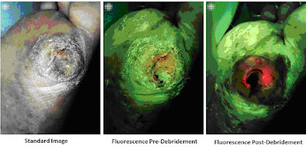
From a 2019 study (by Raizman et al) about use of this technology (MolecuLight i:X) with debridement, scans of 22 wounds were conducted under standard and violet light in patients after debridement. Scans, performed non-invasively at the point-of-care, demonstrated remaining bacteria/biofilm signal in 100% of wounds after sharp debridement, triggering additional removal of tissue. [1] 

Re-scanning demonstrated a marked decrease or complete removal of bacteria in most wounds, yet a subset showed persistent or increased bacterial signals post debridement. The authors, aided by the knowledge gained from this technology,  proposed “the subgroup with persistent bacterial fluorescence post-debridement was at increased risk of deep compartment infection and required more frequent debridement and/or antibiotics.” [1]. See example of this in figure 2 (tissue in green, bacteria in red). Others have demonstrated ability to markedly decrease antibiotic usage using this technology, showing great potential for stewardship efforts [2].

Currently, the technology can take direct images of the skin or mucosal surface to indicate bacteria on the tongue or oral lining. Exploratory reviews of this imaging device also shows possible benefits in scanning potential biopsy material with violet light at the tissue and see if it's suggestive for cancer or inflammatory disease. 


SCAN 1: The Test Drive

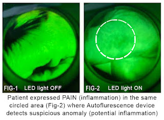
Autofluorescence has been proven and used worldwide to show inflammatory changes and bacterial infection. It's used to find both Gram‐negative bacteria and Gram‐positive bacteria, aerobic and anaerobic. (Fig‐1) The illustrated scan shows the normal skinfolds as green and the skin image is homogeneous (there's nothing dark). (Fig‐2) The scan of the finger on the opposite hand which the patient expressed chronic irritation. Upon initial observationit appears that Autofluorescence may detect inflammation through the upper dermis, a change documented visually and by ultrasound imaging as minimal epidermal thickening associated with inflammatory skin disease (mild type). When we turned on the fluorescence scan, the exact one centimeter area of redness on the skin corresponded to the one centimeter darkened area on the finger.


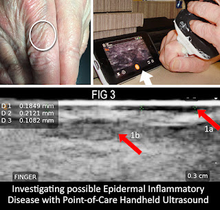
SCAN 2: CLINICAL IMAGING & OBJECTIVE DATA MEASURING WITH ULTRASOUND 
(Fig-3) For my diagnostic research projects in dermal lesions and infections, one of my "weapons of choice" is the doppler ultrasound.  From tumors to traumas, radiologists and clinicians rely on feature-rich computerized systems that provided remarkable patient data for its non‐invasive and real-time view of inflammatory disease to align with our study of the Autofluorescence for its work on atopic dermatitis and other viral diseases (ie. Covid-19) which may affect the skin and be sampled with this noninvasive technology. Since it hasn't been clinically documented in the dermal tissues, this is an opportunity to form a new type of imaging that is non-invasive to consider avoiding biopsies on children.

In the case of Fig-3, I used a high-frequency setting on a popular handheld point of care ultrasound probe to recognize a 0.3cm depth scan of the same area of the finger that was previously scanned by the fluorescence device. The top right arrow (1a) shows the enlarging dark stripe indicating inflammatory thickening or the epidermis. The middle grey area (arrow 1b) represents subdermal inflammation of subcutaneous tissue or mild inflammatory skin disease -  confirming the prior evaluation.

EXPANSION POTENTIAL: RESEARCH 
Fluorescence imaging is proven to have a four‐millimeter depth of diagnostic accuracy in the tongue or mucous membranes‐ similar to the cervix or the vagina or the intestine lining. There is a rich literature of its use in skin wounds and wound infection. However, it's use in skin diseases has not been fully explored‐ so this is a beta test to see if and how it will work in non‐invasive diagnosis of inflammatory skin disease, especially in children. Exploring this technology is of great importance because dermatitis is recognized as a major pediatric disorder. Also disabling inflammatory chronic skin disease (psoriasis, rosacea) are a major focus of successful treatment by pharmacology companies.


AUTOFLUORESCENCE FIELD TEST   
 
by: Randall Weisel, DDS (Excerpt from Journal of Dentistry and Oral Sciences/ "Auto Fluorescence Allows Us to Detect Early Signs of Oral Cancer and Much More")

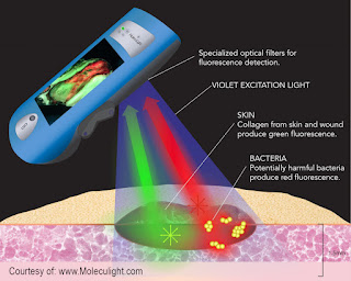
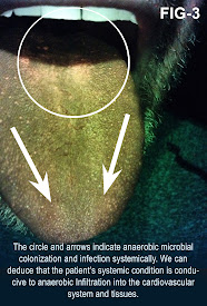
Review of autofluorescence enables us to objectively examine the oral cavity for inflammation and infection. Many systemic diseases are perpetuated by microorganisms that colonize in the oral environment. They enter the cardiovascular system by enzymatic processes that open the oral mucosa to allow their entry. A majority of the microbes are anaerobes and/or facultative anaerobes. When they enter the host, they metabolize blood. Their waste by products contains iron elements within a compound called porphyrin. Porphyrin will fluoresce when exposed to certain wavelengths of light. Healthcare providers can utilize this natural occurring process to objectively see these harmful pathogens. This may indicate that the host has a Sleep Related Breathing Disorder (SRBD). Sleep apnea is a primary disorder of SRBD’s. This technology offers medical and dental fields a screening tool for a pandemic healthcare problem.

Pathogenic microbial presences and the size of the colony (the bioload) can be relatively determined [19]. Fluorescence visualization of red-orange means bacterial pathogens are present. Presence of bluish green/bright white indicates presence of Pseudomonas [19]. The conditions for these pathogens to exist can occur within individuals with Sleep Related Breathing Disorders (SRBD). Sleep Apnea is the most prevalent of these disorders [20]. Researchers should focus their attention to the dorsum of the tongue ecosystem. Autofluorescence, performed with technology that utilizes electromagnetic spectral ranges and special filters that are components of the Velscope, allows visualization of pathogenic microbes. Scientific research demonstrates that oral pathogens (Pg, Aa, and Fn) may be direct and/or indirect causative agents of systemic diseases. These oral pathogens cause inflammation and infection.


SPECIAL THANKS to Dr. Monique Rennie, Director of Scientific Affairs and Global Engagement at Moleculight for her generous technical support and assistance greatly added  to the quality and integrity of this feature article. 


REFERENCES:

1) Raizman R et al. Use of a bacterial fluorescence imaging device: wound measurement, bacterial detection and targeted debridement. J Wound Care (2019). https://pubmed.ncbi.nlm.nih.gov/31825778/

2) Price N. Routine fluorescence imaging to detect wound bacteria reduces antibiotic use and antimicrobial dressing expenditure while improving healing rates: retrospective analysis of 229 foot ulcers. Diagnostics 2020;10:927.

19) Le, L.; Baer, M.; Briggs, P.; Bullock, N.; Cole, W.; DiMarco, D.; Hamil, R.; Harrell, K.; Kasper, M.; Li, W.; et al. Diagnostic Accuracy of Point-of-Care Fluorescence Imaging for the Detection of Bacterial Burden in Wounds: Results from the 350-Patient Fluorescence Imaging Assessment and Guidance Trial. Advances in Wound Care. 2021. https://pubmed.ncbi.nlm.nih.gov/32870774/

20) Sleep-Disordered Breathing.chapter 23.Thoracic.org. 237-247. Boillot A, Demmer RT, Mallat Z, Sacco RL, Jacobs DR, Benessiano J, et al. Periodontal microbiota and phospholipases: the oral infections and vascular disease epidemiology study (INVEST). Atherosclerosis. 2015;242(2):418-23. PubMed | CrossRef

21) Bale BF, Doneen AL, Vigerust DJ. High-risk periodontal pathogens contribute to the pathogenesis of atherosclerosis. Postgrad Med J. 2017;93(1098):215-20. PubMed | CrossRef

22. Boillot A, Demmer RT, Mallat Z, Sacco RL, Jacobs DR, Benessiano J, et al. Periodontal microbiota and phospholipases: the oral infections and vascular disease epidemiology study (INVEST). Atherosclerosis. 2015;242(2):418-23. PubMed | CrossRef

23) Desvarieux M, Demmer RT, Jacobs Jr DR, Rundek T, Boden-Albala B, Sacco RL, et al. Periodontal bacteria and hypertension: the oral infections and vascular disease epidemiology study (INVEST). J Hypertens. 2010;28(7):1413. PubMed | CrossRef


Disclaimer & Copyright Notice: This article/website does not provide medical advice. The information, including, but not limited to text, graphics, images and other material contained on this blogsite are for informational purposes only. No material on this site is intended to be a substitute for professional medical advice, diagnosis or treatment. Furthermore, any/all contributors mentioned in this article are presenting only ANECDOTAL findings of the effects of the products/technologies they are featuring- and are not offering clinical data or medical recommendations in any way. Always seek the advice of your physician or other qualified health care provider with any questions you may have regarding a medical condition or treatment and before undertaking a new health care regimen, never disregard professional medical advice or delay in seeking it because of something you read on this page, article, blog or website.

The materials provided on this website/web-based article are copyrighted 2021 and the intellectual property of the publishers/producers (The NY Cancer Resource Alliance/IntermediaWorx inc. and Bard Diagnostic Research & Educational Programs). It is provided publicly strictly for informational purposes within non-commercial use and not for purposes of resale, distribution, public display or performance. Unless otherwise indicated on this web based page, sharing, re-posting, re-publishing of this work is strictly prohibited without due permission from the publishers.  Also, certain content may be licensed from third-parties. The licenses for some of this Content may contain additional terms. When such Content licenses contain additional terms, we will make these terms available to you on those pages (which his incorporated herein by reference).The publishers/producers of this site and its contents such as videos, graphics, text, and other materials published are not intended to be a substitute for professional medical advice, diagnosis, or treatment. For any questions you may have regarding a medical condition, please always seek the advice of your physician or a qualified health provider. Do not postpone or disregard any professional medical advice over something you may have seen or read on this website. If you think you may have a medical emergency, call your doctor or 9-1-1 immediately.  This website does not support, endorse or recommend any specific products, tests, physicians, procedures, treatment opinions or other information that may be mentioned on this site. Referencing any content or information seen or published in this website or shared by other visitors of this website is solely at your own risk. The publishers/producers of this Internet web site reserves the right, at its sole discretion, to modify, disable access to, or discontinue, temporarily or permanently, all or any part of this Internet web site or any information contained thereon without liability or notice to you.


Friday, November 26, 2021

TODAY’S CLINICAL AESTHETICS- SCIENCE AND STRATEGY


REVIEW OF AESTHETIC DERMATOLOGY
By:  Michelle P. Zappas DNP, FNP

I am a family nurse practitioner for the past 10 years. But my main expertise (and where I work clinically) is in aesthetics or aesthetic dermatology. I currently work in Luxe Aesthetics Center as a clinician or as a family nurse practitioner in Yorba Linda, CA. The CEO of The Luxe Aesthetic Center who is an awesome colleague and mentor in clinical aesthetics. So we do a lot with Botox, Neurotoxins, Fillers laser techniques to bring enhancements to patients, helping them to look and feel their best.

Thanks to the connective powers of LINKEDIN, I was approached by NY Cancer Imaging Specialist, Dr. Robert L. Bard about co-authoring in a multi-disciplinary text project about IMAGE GUIDED AESTHETIC PROCEDURES.  Having learned about him as a globally recognized figure in clinical education and medical publishing,  I also marveled at his vision to unite the many areas of medicine through collaborative research and integrative presentation concepts.  Meanwhile, to co-write about clinical aesthetics in this unique capacity was a remarkable opportunity no educator in my field could pass up.

INSIGHTS ON SAFETY
 So when doing hyaluronic acid filler, it is really important to know which areas are dangerous. Then it basically breaks down to nerves and arteries. Obviously you don't want to occlude an artery or with filler itself or too much filler into an area where that presses on an artery, because that can cause vascular compromise avascular necrosis, it can cause blindness if the artery supplies the eye with blood and also you can cause a palsy if there is occlusion or compression of the nerve.

 So those are really the major vessels and anatomic areas to avoid-- the, the big subgroups.  But obviously within the face, there are a lot of vessels and arteries to note and to avoid when you are injecting. So this chapter really breaks it down. We go through the face upper face, lower face in terms of where to inject safely, what vessels and arteries that are in that area and what to avoid- plus other clinical techniques in order to make sure that you are injecting the hyaluronic acid safely.

I am definitely interested in public education, informing patients and all providers about best practices with regard to aesthetics, other techniques and modalities.  I’d like to contribute in informing the public about aesthetics before getting these procedures, especially in support of the many patients of enhancement or regenerative professionals.  I’d also like to promote safety programs and prevention techniques because a lot of damage and injury can happen out there, particularly with fillers but also with lasers if it's not done correctly. So it's really important when we're talking about patient safety for the provider to be able to walk the line between being aggressive enough to get somebody good results but (especially in terms of lasers) not work too aggressively to burn somebody or to cause somebody scars or blisters.

IPL- INTENSE PULSE LIGHT THERAPY
 
So Intense pulse, light or IPL is a type of laser that can target the main chromophores (hemoglobin, water, and melanin) and so these are a lot of the molecules that patients complained about in chunks of  dyschromia or erythema on their face and other parts of their body. A lot of times people are coming into decrease the Pohnpeian with the little blood vessels in the skin. IPL is wonderful for that.

One of my points about my IPL chapter is that it's not just for aesthetics; there are a lot of dermatologic inflammatory.  Multi-system conditions and diseases that IPL can be used for include ROSACEA, Meibomian Gland dysfunction, Acne,  Port-Wine Stains related to Sturge–Weber syndrome and Mangiomas. There are a wide variety of conditions that can cause patients discomfort- including mental discomfort,  suicidal ideation and  depression that can be treated and a radiated with IPL as one of the modalities for treatment.  Hence, I find this to be a remarkable laser for a lot of different health conditions while also strongly making a significant difference in the areas of aesthetics as well as dermatologic conditions.


In terms of our fillers, using image guidance via ultrasound, we can visualize vessels, nerves (and) all sorts of different things that allow the practitioner to safely inject. And so that is a great technique to not occlude those vessels that you don't want to occlude. To transform somebody's face and give them a youthful appearance, but safely without the risk of blindness or vascular necrosis or any other of the potential adverse effects that can come from that kind of hyaluronic acid or other filling modalities.

ON ANTI-AGING AND REGENERATION
 
Clinical aesthetic modalities as a whole works on the visual of the patient but also greatly improving their mental wellbeing and their ability to interact with the world (resulting to improved self-esteem). And I don't want to get lost on the vanity aspect of it because aesthetics actually running a whole lot deeper than that. My philosophy behind that is to “do what the patient wants” in terms of what bothers them.  However, I do not buy into all of these new modalities necessarily without seeing the evidence. I really want the evidence to support the procedure. So patients who invest in these modalities deserve my research, my clinical understanding and I want to make sure that what I am providing them that is backed by evidence. So that is probably where I talk some patients out of some of the newer things that they see in the blogosphere and YouTube from the many influencers- and I say, “let's go back to what has been tested, where we can have the evidence—that’s what makes for a sound and professional clinical aesthetics practice!”


.

EXTRA:

Thwarting Surprise "Surgical Land Mines" with PreOp Scans

By. Dr. Robert L. Bard

For all my friends in the practice of Cosmetic Surgery, DIGITAL PRE-OP is a highly useful stage for many patients who may carry hidden issues that can turn into a pandora's box of complications. I have performed this vital service for European plastic surgeons since 2001 in their centers  while currently performing domestically as a digital diagnostics partner for serious physicians and surgeons fulfilling similar needs.  Pre-operative imaging is widely performed to verify tissue planes and measure fat depth.  Since patients may have forgotten prior treatments, new scans sometimes reveal extensive sub-dermal calcium deposition, unsuspected fluid collections or thick fibrosis distorting the expected anatomy.  Anatomic variants may be observed and avoided. Moreover, patient confidence is enhanced by the extra care provided by this advanced technology.

Some of the most common POST-PROCEDURE COMPLICATIONS include:
- Suture loosening and granuloma formation following blepharoplasty
- Lipoatrophy or fat necrosis following PRP or abdominoplasty
- Filler complications and implant migration 

- Doppler verification of vascular compromise (venous or arterial) following facial therapies allowing immediate intervention to prevent blindness or tissue necrosis (See complete article)


TEXTBOOK OVERVIEW: 
Springer Medical Publishing proudly presents the first installment in clinical aesthetic procedures. This detailed and up-to-date overview of image-guided procedures focuses on the many aesthetic and reconstructive strategies delivered by some of today's renowned leaders in the clinical aesthetics community. They share their valuable expertise and field-based findings throughout this feature-rich textbook. The wide list of audiences for this text (ie. dermatologists, plastic surgeons, aestheticians, general surgeons) will enjoy an insider's look at each treatment program while providing remarkable field-based knowledge for the general non-medical audience seeking the latest information in non-invasive and minimally invasive aesthetic procedures.  Produced and edited by Dr. Robert L. Bard, this collective project showcases the most highly sought-after cosmetic treatments in each priceless chapter- through detailed breakdowns, experiential insights and a generous graphic tour of before and after progress visuals.  Thanks to the additional safety benefits of clinical imaging, our treatment professionals express added confidence in the pre-operative and post-op areas. In addition, many aesthetics procedures noted also brings significant advantages (of accuracy and efficiency) to the actual treatment process from real-time image guidance. Some of our top contributors include: Dr. Beth Haney, Dr. Michelle Peters-Zappas, Mary Nielsen Aesthetics, Dr. Arun Garg, Dr. John Catanzaro, Dr. Randall Weisel, Dr. Stephen Chagares, Dr. Cari Green, Dr. Richard Kushner, Dr. Lio Yu, Dr. Peter A Everts and January Howard, CMA (this is an abridged list of our contributors).


ABOUT THE AUTHOR

Dr. Michelle (Shelly) Zappas is a Clinical Associate Professor at the USC Suzanne Dworak-Peck School of Social Work in the Department of Nursing. She received her Bachelors’, Masters’ and Doctorate in nursing from Columbia University. Dr. Zappas has nearly a decade of experience as a family nurse practitioner in primary care clinics in New York City and Los Angeles. Dr. Zappas has authored multiple publications and presents at both state and national nurse practitioner educational conferences. In aesthetics she has been trained in advanced injection techniques using neurotoxins and dermal fillers as well as training in IPL and lasers. Dr. Zappas has a profound interest in dermatology and skin care and enjoys helping her patients put their best face forward.  


Disclaimer: The information (including, but not limited to text, graphics, images and other material) contained in this article is for informational purposes only. No material on this site is intended to be a substitute for professional medical advice or scientific claims. Furthermore, any/all contributors (both medical and non-medical) featured in this article are presenting only ANECDOTAL findings pertaining to the effects and performance of the products/technologies being reviewed - and are not offering clinical data or medical recommendations in any way. Always seek the advice of your physician or other qualified health care provider with any questions you may have regarding a medical condition or treatment and before undertaking a new health care regimen, never disregard professional medical advice or delay in seeking it because of something you read on this page, article, blog or website.


Monday, October 4, 2021

MEDTECH REVIEWS 2: Introducing TMS (Transcranial Magnetic Stimulation)

The Modern Age of Non-Invasive Mental Health Innovations: Transcranial Magnetic Stimulation
Written By: Jessica Connell-Glynn | Dr. Robert L. Bard & the editorial team of prevention101.org

In the continued expansion of the medical society to “go non-invasive” (or non-surgical) and the trending reassessment of interventional medications, the mental health community has acquired major tools in its toolbox to support clinical diagnostic and treatment efforts.  In 2008, the FDA approved the first TMS Depression Device for Depression Treatment [1]. In 2018, the FDA approved the marketing of Repeated Transcranial Magnetic Stimulation (rTMS) as adjunct therapy for of obsessive compulsive disorder (OCD) [2-3].  

Current reports have presented an est. 30% of depression cases have a resistance to antidepressant drugs, where Transcranial Magnetic Stimulation (TMS) and the application of Transcranially applied non-invasive neuro-magnetic intervention has shown positive results in combination with antidepressants in patients with treatment-resistant depression[ 4]. 

During the procedure, a magnetic coil is positioned at the head of the person receiving the treatment using anatomical landmarks on the skull, in particular the inion and nasion.[5] The coil is then connected to a pulse generator, or stimulator, that delivers electric current to the coil.[6] Single or paired pulse TMS causes neurons in the neocortex under the site of stimulation to depolarize and discharge an action potential. [7] Repetitive TMS produces longer-lasting effects which persist past the period of stimulation. rTMS can increase or decrease the excitability of the corticospinal tract depending on the intensity of stimulation, coil orientation, and frequency. [8]

Field Report: TMS Science from the Top Developers
Based on an interview with Nexstim Vice President, STEVE BELLER

TMS, or transcranial magnetic stimulation, is the use of magnets external to the body to activate tissue inside the body (so you're not having to open the patient up).  Based on Faraday’s Law, a magnetic field produced outside of a patient’s head can permeate non-invasively through a patient’s head and induce an electric field that has the capacity to activate neurons in the brain. We induce current at a distance inside the brain and cause the neurons to fire where we induce that current.  This means that where we depolarize, we cause the brains neurons to fire.  TMS artificially stimulates the brain and causes the neurons to fire.  Navigation technology allows us to see precisely where in the brain where we are stimulating.

The value of this stimulation effect is twofold; the first is DIAGNOSTIC- where we can non-invasively identify specific areas of motor function and specific areas of language function. The second function is THERAPEUTIC- where we can actually help change or fix an identified brain disease by using TMS. Right now,  we as a company hold FDA clearances for pre-operative motor and language mapping as well as treating major depressive disorder. For depression: the general understanding (still under exploration like a lot of things in medicine, particularly with the brain) is that the metabolic activity in the left dorsal lateral prefrontal cortex in a diseased or depressed patient is lower and usually significantly lower than in out of a healthy patient. In order to increase the metabolic activity and the left dorsal lateral prefrontal cortex or left DLPFC you can use TMS to artificially stimulate our cause the neurons to fire in the left DLPFC and that increases the neuronal excitability level and helps improve the depressive state of that patient.  This is one example of how to therapeutically use TMS to improve health and performance. 

You can also use a different set of TMS stimulation parameters to inhibit brain regions that may be overactive. That is, you can actually slow down the functioning of the cortical area that you're targeting. Hence, TMS in therapeutics may be a versatile option to treat a range of neurologic and psychiatric disorders in the future.


EXTRA:

Navigated TMS for Pre-Procedural Brain Surgery

Since 2009, Nexstim´s Navigated Brain Stimulation (NBS) has been FDA cleared for use in non‐invasive motor and language functional cortical mapping prior to radiosurgery and invasive neurosurgery. This technology allows the operator to move a hand‐held TMS coil over the patient´s head delivering a series of harmless magnetic pulses whose electric fields gently activate neurons in the cortex. Converting a standard head MRI into a 3D‐model of the patient´s brain, allows the operator to precisely navigate the coil´s electric field anywhere in the cortex, much like driving a car with the aid of GPS. Measuring patient responses by electromyography (EMG) reveals the locations of the vital motor areas inside the brain. Mitchel Berger, M.D, Past Chairman of the Department of Neurological Surgery, Past Director of the Brain Tumor Surgery Program, University of California, San Francisco, said, “What makes this so special is the ability for it to define very small, precise cortical regions on the surface of the brain, and define individual components of function which is something we never could do before." The Nexstim technology is available to patients at over 50 of the country’s top neurosurgery programs.


2021 REVIEW OF POST TRAUMATIC RECALL
 (A field report by: Jessica Connell-Glynn, LCSW)
 PTSD can occur in all different extremes with at-risk professionals (like cops, responders and veterans). The trauma that they experience are above the ordinary that they could cause extreme flashbacks, anxiety and depression—heavily affecting their quality of life. The average civilian is also prone to this disorder starting with MICRO-TRAUMAS that can happen to everybody throughout any point in their lifetime.  Usually stemmed from childhood issues, micro-traumas actually shape the way an individual reacts to other people. As an example, child bullying may lead to developing a protective or defensive personality disorder.  Anytime they feel disrespected or embarrassed by others, feelings of extreme uncontrollable anger may arise without knowing the source of the hurt or why they're acting in that way.  This dilemma often causes problems in relationships.


LANGUISHING: EMOTIONAL CRISIS FROM THE PANDEMIC
 By: Jessica A. Conner-Glynn, LMSW  / Edited by: Cheri Ambrose (NYCRA NEWS writing staff)
 Observing the many emotional after-effects of our ‘new Covid lifestyle’, I find a link between stepping away one's our work environment and a direct improvement in one’s well-being. The surge of the coronavirus pandemic brought a unique kind of widespread emotional disorder called LANGUISHING- an issue identified as somewhere between a downturn ‘blah’, emptiness or depression causing a type of burnout from mandatory social distancing.  Many of us are working really hard in the same place that we actually live- removing that safe separation between work and home that we once had.  The shift in working at home takes away that place to relax and shut down, eventually causing a state of emotional exhaustion from staying ON (and on guard) the whole time.  There is also a physical aspect to all this; the new sedentary lifestyle of working from home eliminates the vital energy stimulation from circulation and cardiovascular activity of physical traveling.  In addition, self-isolating has induced an unhealthy weight gain in many, also adding to this malaise.


HOW STRESS CAN MAKE YOU SICK By: Dr. Jesse A. Stoff 
 For our well-being, stress is so powerful that it can affect just about every function in our bodies right down to our cells. Stress can cause major weight gain, headaches, hair loss, dental problems, major fatigue, respiratory issues, digestive problems, psychological & focal disorders.  It is also known to affect the performance of our immune system - allowing for skin issues, colds, allergies and a host of chronic diseases. Stress management is important for more than psychological health; stress can greatly affect immune functioning, inflammation and even hormonal balance. Studies have shown that stress can reduce natural killer cell function. These are normally the cells that attack foreign invaders including viruses, bacteria and fungi. They are also the cells that patrol the body as the first line of defense against the development of cancer.


RETINAL & TRANSCRANIAL IMAGING By Dr. Robert L. Bard
Another device used by imaging specialists to detect mental distress is through an EYE SONOGRAM or  Real Time Sonofluoroscopy of the orbital soft tissues of the eyes.  This process is performed in multiple scan planes with varying transducer configurations and frequencies.  Power and color Doppler use angle 0 degrees and PRF at 0.9 at the optic nerve head. 3D imaging of optic nerve and carotid, central retinal arteries and superficial posterior ciliary arteries performed in erect position before & after verbal communication and orbital muscle tissue contractions may be observed as a precursor to visual changes in facial expression. Retinal arterial directional flow is also measured with peak systolic and diastolic values. Bulging of the optic nerve head is checked as increased intracranial pressure may be demonstrable in this condition. Other innovations such as the TRANSORBITAL DOPPLER, 3D/4D VESSEL DENSITY HISTOGRAM and the RETINAL OCT (optical coherence tomography) are also being explored in the pursuit of studying brain performance through the eyes.  An expanded review on these solutions will be available in part 2 of this report.


References:

1) “FDA OKs TMS Depression Device for Brain-Stimulating Device Cleared for Depression Treatment After 1 Drug Failure”- https://www.webmd.com/depression/news/20081008/fda-oks-tms-depression-device

2) https://www.fda.gov/news-events/press-announcements/fda-permits-marketing-transcranial-magnetic-stimulation-treatment-obsessive-compulsive-disorder

3) https://www.hcplive.com/view/prevalence-methamphetamine-use-overdose-death

4) Biological Psychiatry (Journal) /ARCHIVAL REPORT| VOLUME 68, ISSUE 2, P163-169, JULY 15, 2010 Transcranial Low Voltage Pulsed Electromagnetic Fields in Patients with Treatment-Resistant Depression  https://www.biologicalpsychiatryjournal.com/article/S0006-3223(10)00162-9/fulltext

5) Nauczyciel C, Hellier P, Morandi X, Blestel S, Drapier D, Ferre JC, et al. (April 2011). "Assessment of standard coil positioning in transcranial magnetic stimulation in depression". Psychiatry Research. 186 (2–3): 232–8. doi:10.1016/j.psychres.2010.06.012. PMID 20692709. S2CID 25100990.

6) NICE. January 2014 Transcranial magnetic stimulation for treating and preventing migraine.  Michael Craig Miller for Harvard Health Publications. July 26, 2012 Magnetic stimulation: a new approach to treating depression?

7) Pascual-Leone A, Davey N, Rothwell J, Wassermann EM, Puri BK (2002). Handbook of Transcranial Magnetic Stimulation. London: Edward Arnold. ISBN 978-0-340-72009-7.

8) Fitzgerald PB, Fountain S, Daskalakis ZJ (December 2006). "A comprehensive review of the effects of rTMS on motor cortical excitability and inhibition". Clinical Neurophysiology. 117 (12): 2584–96. doi:10.1016/j.clinph.2006.06.712. PMID 16890483. S2CID 31458874.

9) Wikipedia: Transcranial magnetic stimulation  https://en.wikipedia.org/wiki/Transcranial_magnetic_stimulation#cite_note-Handbook_of_Transcranial_Magnetic_Stimulation-27


Saturday, September 25, 2021

WHAT IS PEMF Therapy? (Pulsed ElectroMagnetic Field)- Part 1

NON-INVASIVE ELECTROMAGNETIC THERAPIES (Part 1) 
A report by: Jessica Connell-Glynn / Edited by the NYCRA-NEWS Editorial team

In 2015, Dr. Oz aired a program about an "invisible treatment" for chronic pain designed to "change the practice of medicine"- casting a wave of commercial attention to the patient community desperate for pain relief- and hope for the reversal of pain-related ailments. Pulsed Elecromagnetic Field Thereapy or PEMF is the induction of electromagnetic fields to promote the synthesis of cellular, muscular and skeletal matrix [1].  

Electromagnetic fields (EMFs) provide a non-invasive, safe, and easy method to treat and rehabilitate a growing list of health issues. The therapeutic theory behind this innovation is designed to affect the body at the extracellular level- imposing the ability to transduce or separate mechanical charge to electrical energy.  This has been reported to affect the alignment and physical properties of tissues & cell nutrition toward the healing process.[2]  Electromagnetic fields (EMFs) and magnetic therapy presents a major increase in research attention in the past decade for its ability to directly treat the site of injury. [3]

Medical historians and publishers often attribute the scientific roots of electrical transduction therapy to NIKOLA TESLA (1856-1943) for his pioneer work in advanced electromagnetic field frequency. They align the success of non-invasive treatment innovations including the Pulsed Elecromagnetic Field Thereapy (PEMF) and the Transcranial Magnetic stimulation (TMS) to the philosophy of implementing electgromagnetic fields to stimulate cells.  It is this same paradigm in patient care technology that enforces the very backbone of holistic and non-surgical ("no more scalpel") movement.


Conventional uses of magnetic therapy is widely found in the integrative and alternative (or wellness) communities. Some may recognize this science under other names including:
 Wave Therapy                   Biofeedback                 Bio-resonance
 Magnetic Therapy             Electrostimulation       Microcurrent Therapy

These devices vary in size & designs but these protocols are similar in application.  The science employs a magnetic field for rehabilitation to treat and/or prevent diseases.

CLEARED FOR USES
In 1979, The Food and Drug Administration 1979 approved PEMF Therapy for the healing of non-union fractures [4]. Electrical stimulation of the spine (as part of spinal fusion procedures) for failed fusions and congenital pseudarthroses. In October 2008 the Food and Drug Administration approved the use of PEMF therapy for treatment of major depressive disorder in PD patients who failed to achieve satisfactory improvement from very high dosages of antidepressant medications [5].  

Clinical research has also been highly dedicated towards mental health. In 2006, the FDA approved PEMF Therapy for treatment of depression and anxiety. [7-9] Further reports have presented an est. 30% of depression cases have a resistance to antidepressant drugs, where Repetitive Transcranial Magnetic Stimulation (rTMS) and the application of Transcranially applied Pulsed Electromagnetic Fields (T-PEMF) has shown positive results in combination with antidepressants in patients with treatment-resistant depression[10].

In 2019 the wearable RecoveryRx PEMF Device was FDA Cleared for "Adjunctive treatment of postoperative pain".  This technology achived market clearance for its drug-free (non-opiod) PostOp effects. Andrew Whelan, President, expressed confidence in seizing the market opportunity: “We are delighted that the FDA has recognized the potential of RecoveryRx in transforming postoperative pain care. RecoveryRx, with its high degree of safety, excellent clinical evidence of effectiveness and exceptional cost-benefit, will become the standard of care.” [11-12]


PEMF: HISTORY & EXPANSION OF MODERN TARGET ISSUES
Claims of electricity induction in aiding bone repair has been reported as far back as the mid 1800's, but it wasn't until the mid-1950's to the '70's that the scientific community significantly advanced its research toward the treatment of delated fractures in Western Europe and the US. Recent reviews have shown benefits of PEMF to include: neurological disorders, vascular disease, lung disorders, GI (gastrointenstinal) disease, rheumatic disease, dermatological issues, inflammation, tumors and immunological issues for both humans and animals.[14] Clinical research studies have reported benefits for a host of specific issues including: musculoskeletal(MSK) Pain [15], bacteria and parasites, OstheoArthritis, mental health disorders (Depression), Multiple Sclerosis[16-17], Cancer[18] and Alzheimer's Disease [19].  

TMS and PEMF USE IN PARKINSON'S DISEASE
Parkinson’s disease (PD) is one of the most common progressive nervous system disorders that affects movement.  It is a neurodegenerative disorder second only to Alzheimer’s disease, accompanied by the impairment of the cortico-subcortical excitation and inhibition systems, hence belonging to the involuntary movement diseases. In the first PD patients treated with high-frequency TMS in 1993, motor symptoms, tremor, rigidity and akinesia improved significantly allowing to decrease the administration of l-dopa by a mean of 55% [20].


TMS FOR DEPRESSION: Transcranial Magnetic Stimulation

October 4, 2021- TMS, or transcranial magnetic stimulation, is the use of magnets external to the body to activate tissue inside the body (so you're not having to open the patient up).  Based on Faraday’s Law, a magnetic field produced outside of a patient’s head can permeate non-invasively through a patient’s head and induce an electric field that has the capacity to activate neurons in the brain. We induce current at a distance inside the brain and cause the neurons to fire where we induce that current.  This means that where we depolarize, we cause the brains neurons to fire.  TMS artificially stimulates the brain and causes the neurons to fire.  Navigation technology allows us to see precisely where in the brain where we are stimulating. (see complete article)


The AOPP: Explaining How PEMF Works


9/25/2021:  An exploratory review* of the PEMF science is under discussion between Dr. Robert Bard and THE AOPP FOUNDATION (Association of PEMF Professionals) which aims to provide clarity on the science behind its 'invisible healing' concept.  AOPP aligned with the Integrative Pain Healers Alliance (Dr. Bard's wellness network) and saw the need for public clarity in how PEMF works, what actual uses they are cleared for as well as the importance of public guidance on the market.

Dr. Jerry Dreesen, President of the AOPP and certified chiropractor by trade is an avid user of this therapeutic technology for his patients on a variety of disorders.  "So PEMF did start out on the animal side... it was originally used for seed germination -and moved over to the animal side.  When they eventually started working with humans, evidence of creating better blood flow as part of the body's detoxification. We're also finding that with PEMF is that the body is utilizing medication even better -  so it's absorbing it and using it more versus some people can be getting a reaction to better absorption of their medications."

Dr. Dreesen further explains the physiological and biological reactions to electromagnetic science and its research - going as far back as the Krebs cycle (otherwise known as The citric acid cycle (CAC) – or  the TCA cycle (tricarboxylic acid cycle)- which we all had to learn. During that process. one of the by products is nitric oxide and then the other one is of course, taking ADP and attaching the phosphate to it and making ATP-- creating the "fuel" for the body.  PEMF sparking this process is great especially for sports where it induces renewed energy to fatigued muscles. For the same reasons why it works really well on animals, the same goes for humans after a workout as it quickly revitalizes the target area by increasing absorption of oxygen and energy levels. 

Dr. Dreesen shares the rehabilitative theories and applications for MSK injuries- including sprains, strains, joint issues, arthritis pain and other chronic inflammation. He describes its performance on a cellular level and controlling inflammation versus "just telling the brain that there's no pain there at all... it's not masking pain, it's physically going in and eliminating the creation of inflammation. As a by-product of going in and oxygenating tissue, creating stem cells, eliminating causing the cellular walls to get rid of product and bringing in nutrients, it rebuilds and refreshes the joints."   He furthers, "for people who have had past injuries in their joints and for whatever reason, never healed properly, PEMF can offer significant change- not only in reducing inflammation, but potentially preventing it from coming back!" 

MEETING DEVELOPERS
Tech critic and test-driver Dr. Robert Bard met with a short list of  AOPP members including Patrick Ziemer, development facilitator and CTO of Aura Wellness PEMF (Aurawell.com) to discuss the growing number of health issues and disorders that neuromagnetic science is slated to manage, treat and even resolve.  As part of his global crusade to support non-invasive medicine, Dr. Bard recognizes the advancing benefits of PEMF throughout the wellness community and is evaluating all devices including the AuraPulse™ technology. 

Mr. Ziemer is a consumer advocate for the AOPP and publishes educational posts about the benefits and his evidence of the PEMF technology to align with his critical thoughts on the industry.  From our interview, he shares his expansive research on the science, allotting for the limited number of actual models that are FDA cleared, approved or registered.   As part of his advocacy, Mr. Ziemer describes the confusion about neuromagnetic therapy to come from companies who presents “rogue claims” about their specific devices.  Many lower-end companies tend to lack the proper extensive safety testing of their devices and espouse claims “from studies done by others that are FDA approved.  For example, companies will make claims that their devices will heal broken bones.  "PEMF has been known to do this, but we can't legally say that about OUR specific device without doing the studies (ourselves) from a formal FDA 510K clearance process … and all companies should take the same responsibility about their work”.  

Mr. Ziemer’s history with PEMF started around 2013, co-developing and promoting the therapeutic use high-voltage PEMF for the equestrian society.  His device appealed greatly to the veterinary community for its ability to address the many health concerns of our four legged friends who rarely complain.  Therapeutically, his PEMF has shown to improve horses’ circulation, increasing blood oxygen, lymphatic stimulation, relaxing muscle spasms, relieving tension, enhancing muscle tone, and increasing the range of motion in high performance horses.  These same health issues expanded his popularity to treat a growing range of patient breeds including cattle, livestock and dogs across the country.

In 2020, Mr. Ziemer’s PEMF officially made its way to human patients at the launch of Aura Wellness- carrying a professional grade, high voltage PEMF device. Applying much of his field expertise with animals, Mr. Ziemer achieved tremendous R&D advantage to apply to his human prototype. His AuraPulse™ model is completely programmable with adjustable intensity levels, digital timers, plug-and-play applicators, and customized session programs.   Dr. Bard is reviewing* all PEMF devices like the AuraPulse™ for its effects and claims for improved wellness.

"Our first test subject was 4 legged and suffered from arthritis. While on the magnetic coil, the poodle visibly became calmer, eventually going to sleep inside the treatment ring. The dog’s handler saw the improvement in the gait and asked to have her shoulder treated. After a 10 minute session, our 2 legged patient reported mild improvement on a chronic rotator cuff disorder and great improvement 12 hours later." 

*It is to be noted that beta testing and clinical evaluation reviews of all medical devices are restricted to their intended uses according to their designated FDA clearances. 


CONTRIBUTORS

JESSICA CONNELL-GLYNN, LCSW, CPC, CEC is a columnist for Prevention101 newsletter. She often reports on topics concerning mental health, neurological disorders and medical innovations that pertain to 'modern wellness'.   As a therapist and mental health coach, Jessica's primary work involves providing direct support in managing symptoms of personal anxiety and panic. Jessica is also currently leading an interdisciplinary research project with a team of mental health and professionals in neurology to review the current care standards and collaborate on new protocols that target, assess and diagnose psychological disorders.(https://cmpcnyc.com/)

Dr. JERRY DREESSEN is both Trauma Team Qualified and Hospital Qualified treating chiropractor in Mountlake Terrace, WA. He is the Executive Director for the Association of PEMF Professionals- the largest professional PEMF organization in the United States. AOPP attracts the most principled and accomplished PEMF user- setting standards by providing certification for all PEMF users, as well as Continuing Education (CEU's) for all members to maintain best practices and techniques. (www.backtoaction.com)

SPECIAL THANKS
The editors of this spotlight proudly gives thanks to Ms. Suzanne Wheeler for her generosity in sharing her story and her resources with us.  Additional thanks to Dr. Jerry Dreessen of the AOPP (Association of PEMF Professionals) and Pat Ziemer of Magnawave Inc. and Aura Wellness PEMF for coordinating our interviews, shared countless materials and conducted unending support to help our educational program bring new light to PEMF technology for chronic disorders and supportive testimonials in alternative therapeutics.   


VIEWPOINTS from the Healthcare Community

Our bodies are made up of 30 to 40 trillion cells at any given point during our lifetime. Each cell is a dynamic electro chemical entity that work together for the better of the whole. This is truly an amazing process. Medicine has been oriented toward rescue type interventions of dysfunction and disease. If there is pathology present;” take it out”. Or use a drug to turn on or turn off the metabolic machinery of specific cells. That is the majority of the mindset of medicine.

It is refreshing to learn about other forward thinking modalities and innovations in the healthcare field. These  Innovations like pulsed electromagnetic field therapy(PEMF) and transcranial magnetic stimulation (TMS) are technologies that utilize non-invasive electromagnetic stimulation of cellular metabolic processes within various places in our body.  The endgame of the technologies is for diagnostic information and therapeutic intervention of systemic disease.  PEMF and TMS have proven that electromagnetic fields can provide non-invasive methods to diagnose and treat health concerns such as depression, pain, MS, Cancer, Alzheimer’s Disease, Parkinson’s Disease, healing responses, and Overall cardiovascular anomalies. This is so exciting because we can use this technology two recognize early metabolic and possibly genetic changes that could lead to accelerated aging disease and death. Also, we can use this technology as an intervention to the changes and correct the path of health. Both of these technologies have shown efficacy ( FDA approval) and promise ( more research is being done). There is no question that LESS IS MORE! That is if we recognize pathological conditions early and can correct them with little or no pharmacological or surgical intervention; “everybody wins!”  


- By Randall Weisel DDS,MPS  (www.laserhygienics.com)          


REFERENCES
1) Ehnert, Sabrina; Schröter, Steffen; Aspera-Werz, Romina H.; Eisler, Wiebke; Falldorf, Karsten; Ronniger, Michael; Nussler, Andreas K. (December 2019). "Translational Insights into Extremely Low Frequency Pulsed Electromagnetic Fields (ELF-PEMFs) for Bone Regeneration after Trauma and Orthopedic Surgery". Journal of Clinical Medicine. 8 (12): 2028. doi:10.3390/jcm8122028. PMC 6947624. PMID 31756999.


2) ACCELERATION OF FRACTURE REPAIR BY ELECTROMAGNETIC FIELDS. A SURGICALLY NONINVASIVE METHOD: First published: October 1974 https://doi.org/10.1111/j.1749-6632.1974.tb26794.x

3) Electromagnetic Field Therapy: A Rehabilitative Perspective in the Management of Musculoskeletal Pain – A Systematic Review J Pain Res. 2020; 13: 1385–1400. Published online 2020 Jun 12. doi: 10.2147/JPR.S231778  PMCID: PMC7297361 PMID: 32606905 https://www.ncbi.nlm.nih.gov/pmc/articles/PMC7297361/


4) Electrical stimulation of the spine as an adjunct to spinal fusion procedures". Blue Cross & Blue Shield of Mississippi. Archived from the original on 2015-04-02. Pulsed electromagnetic field systems with FDA PMA include the EBI Bone Healing System from Electrobiology, Inc., which was first approved in 1979 and indicated for nonunions, failed fusions, and congenital pseudarthroses; and the Cervical-Stim from Orthofix, which was approved in 2004 as an adjunct to cervical fusion surgery in patients at high risk for non-fusion. https://web.archive.org/web/20150402122818/http://www.bcbsms.com/index.php/index.php?id=200&action=viewPolicy&path=%2Fpolicy%2Femed%2FElectrical+Stimulation+of+the+Spine+as+an+Adjunct+to+Spinal+Fusion+Procedures.html&source=emed


5) Mechanisms and therapeutic applications of electromagnetic therapy in Parkinson’s disease (BMC/SPRINGER) Behavioral and Brain Functions-
https://www.ncbi.nlm.nih.gov/pmc/articles/PMC4562205/#CR125

6) Clinical significance of transcranial magnetic stimulation (TMS) in the treatment of pharmacoresistant depression: synthesis of recent data.
Demitrack MA, Thase ME  Psychopharmacol Bull. 2009; 42(2):5-38. https://pubmed.ncbi.nlm.nih.gov/19629020/

7) Pulsed Electro Magnetic Fields (PEMF) in Depression (PEMF)  https://clinicaltrials.gov/ct2/show/NCT03556735

8) Demitrack MA, Thase ME. Clinical significance of transcranial magnetic stimulation (TMS) in the treatment of pharmacoresistant depression: synthesis of recent data. Psychopharmacol Bull. 2009;42(2):5–38 - https://pubmed.ncbi.nlm.nih.gov/19629020/

9) Effectiveness of transcranial magnetic stimulation in clinical practice post-FDA approval in the United States: results observed with the first 100 consecutive cases of depression at an academic medical center- https://pubmed.ncbi.nlm.nih.gov/22579164/


10) Biological Psychiatry (Journal) /ARCHIVAL REPORT| VOLUME 68, ISSUE 2, P163-169, JULY 15, 2010 Transcranial Low Voltage Pulsed Electromagnetic Fields in Patients with Treatment-Resistant Depression  https://www.biologicalpsychiatryjournal.com/article/S0006-3223(10)00162-9/fulltext

11) US FDA- https://www.accessdata.fda.gov/cdrh_docs/pdf19/K190251.pdf
12) Press Release/BioElectronics Announces FDA Market Clearance for Its Non-Opioid Postoperative Pain Therapy   https://www.globenewswire.com/en/news-release/2019/07/01/1876675/0/en/BioElectronics-Announces-FDA-Market-Clearance-for-Its-Non-Opioid-Postoperative-Pain-Therapy.html

13) 12/10/2009 -FDA clears Nexstim´s Navigated Brain Stimulation for non-invasive cortical mapping prior to neurosurgery (Nexstim Press release)
https://nexstim.com/news-and-events/news/press-release/news/fda-clears-nexstims-navigated-brain-stimulation-for-non-invasive-cortical-mapping-prior-to-neurosur/


14)  Jerabeck, J; Pawluk, W (1998). Magnetic therapy in eastern Europe : a review of 30 years of research. W. Pawluk. ISBN 0-9664227-0-8.

15) Thomas AW, Graham K, Prato FS, McKay J, Forster PM, Moulin DE, et al. A randomized, double-blind, placebo-controlled clinical trial using a low-frequency magnetic field in the treatment of musculoskeletal chronic pain. Pain Res Manage J Can Pain Soc (journal de la societe canadienne pour le traitement de la douleur) 2007;12(4):249–258 https://www.ncbi.nlm.nih.gov/pmc/articles/PMC2670735/

16)  Lappin MS, Lawrie FW, Richards TL, Kramer ED. Effects of a pulsed electromagnetic therapy on multiple sclerosis fatigue and quality of life: a double-blind, placebo controlled trial. Altern Ther Health Med. 2003;9(4):38–48. https://pubmed.ncbi.nlm.nih.gov/12868251/

17) Richards TL, Lappin MS, Acosta-Urquidi J, Kraft GH, Heide AC, Lawrie FW, et al. Double-blind study of pulsing magnetic field effects on multiple sclerosis. J Altern Complement Med. 1997;3(1):21–29. doi: 10.1089/acm.1997.3.21. https://pubmed.ncbi.nlm.nih.gov/9395691/

18) Barbault A, Costa FP, Bottger B, Munden RF, Bomholt F, Kuster N, et al. Amplitude-modulated electromagnetic fields for the treatment of cancer: discovery of tumor-specific frequencies and assessment of a novel therapeutic approach. J Exp Clin Cancer Res. 2009;28:51. doi: 10.1186/1756-9966-28-51
https://www.ncbi.nlm.nih.gov/pmc/articles/PMC2672058/

19) Arendash GW, Sanchez-Ramos J, Mori T, Mamcarz M, Lin X, Runfeldt M, et al. Electromagnetic field treatment protects against and reverses cognitive impairment in Alzheimer’s disease mice. J Alzheimers Dis. 2010;19(1):191–210. https://pubmed.ncbi.nlm.nih.gov/20061638/

20) Behav Brain Funct. 2015; 11: 26. Published online 2015 Sep 7. doi: 10.1186/s12993-015-0070-z
PMCID: PMC4562205  PMID: 26347217  Mechanisms and therapeutic applications of electromagnetic therapy in Parkinson’s disease Maria VadalĂ , Annamaria Vallelunga, Lucia Palmieri, Beniamino Palmieri, Julio Cesar Morales-Medina, and Tommaso Iannitti corresponding author https://www.ncbi.nlm.nih.gov/pmc/articles/PMC4562205/


PHOTO CREDITS
* Nikola Tesla in or around 1890, when the inventor was in his mid-30s. | Photo is in the public domain. Image courtesy of the Library of Congress. https://www.energy.gov/articles/top-11-things-you-didnt-know-about-nikola-tesla

Disclaimer & Copyright Notice: The materials provided on this website/web-based article are copyrighted 2021 and the intellectual property of the publishers/producers (The NY Cancer Resource Alliance/IntermediaWorx inc. and Bard Diagnostic Research & Educational Programs). It is provided publicly strictly for informational purposes within non-commercial use and not for purposes of resale, distribution, public display or performance. Unless otherwise indicated on this web based page, sharing, re-posting, re-publishing of this work is strictly prohibited without due permission from the publishers.  Also, certain content may be licensed from third-parties. The licenses for some of this Content may contain additional terms. When such Content licenses contain additional terms, we will make these terms available to you on those pages (which his incorporated herein by reference).The publishers/producers of this site and its contents such as videos, graphics, text, and other materials published are not intended to be a substitute for professional medical advice, diagnosis, or treatment. For any questions you may have regarding a medical condition, please always seek the advice of your physician or a qualified health provider. Do not postpone or disregard any professional medical advice over something you may have seen or read on this website. If you think you may have a medical emergency, call your doctor or 9-1-1 immediately.  This website does not support, endorse or recommend any specific products, tests, physicians, procedures, treatment opinions or other information that may be mentioned on this site. Referencing any content or information seen or published in this website or shared by other visitors of this website is solely at your own risk. The publishers/producers of this Internet web site reserves the right, at its sole discretion, to modify, disable access to, or discontinue, temporarily or permanently, all or any part of this Internet web site or any information contained thereon without liability or notice to you.