Tuesday, April 12, 2022

WHAT IS BIOFEEDBACK? PERSONALIZED SELF-REGULATING THERAPY

INTRODUCTION
IPHA's MedTech Reviews program is dedicated to the constant search for the most current advancements in health innovations.  We report, review, beta-test and challenge the efficacy of these inventions for public awareness of what works, and how they work. 

Here are just some of the latest non-invasive therapeutic technologies in our catalog of medical devices:
•  BIOLUMINESCENCE probes
•  RadioFrequency (RF) Therapy


Our next tour in our search for non-invasive health innovations is the science of BIOFEEDBACK—and we start our review with the internationally recognized pioneers from ONDAMED. Uniquely integrated with the features of PEMF/Pulsed Electromagnetic Field Therapeutics, this device offers localized tissue stimulation by induction of microcirculation within tissue. While the ONDAMED is approved by health authorities outside of the U.S. for pain relief, soft tissue injuries, and wound healing, the U.S. FDA regulates health claims to only include treatment for stress and stress related disorders.

An historically recognized electronic device earned its mark in progressive wellness, muscle relaxation and the alleviation of functional medical disorders (Jacobson/Harvard, 1938)- and evolved into the core of self-regulating therapeutics. In the early 1920's, a community of researchers founded the biometric readings as part of the early works of EEG, visceral learning & electromyography as the major functions of the biofeedback paradigm. EEG studies the basic principle that brain processes can be brought under voluntary control. Pursuit of the meditative, alpha dominant states continues to be part of today's EEG neurofeedback movement- supporting applications of brain wave control and the therapeutic signal of the device.[1]

In the 1970's the science of biofeedback dug its heels into the treatment of headaches and migraines, high blood pressure, urinary incontinence and lower back pain - among a list of other functional issues. Other devices and neurodiagnostics identify the use of biofeedback as an effective driver of self-regulating scanners of biodata.

- ELECTROMYOGRAPH (EMG): measures muscle fibers & neuromuscular studies. Neurofeedback may sometimes be used as a non-invasive treatment for ADHD, pain, addiction, anxiety, depression, and other disorders. [2]

- THERMAL FEEDBACK: reads migraine headaches & Raynaud's disease

- GALVANIC SKIN RESPONSE METER (GSR): measures electrical changes in the skin (sympathetic nervous arousal) supportive of psychotherapy & behavior therapy to track anxiety and cognitive/emotional threat response

According to Rolf Binder, chief engineer/inventor of the ONDAMED device and owner of the Ondamed Companies, and Dr. Silvia Binder, CEO of the Ondamed Companies, biofeedback is a mind-body process of electronic signals designed to send-then-receive (feedback) information about a person's physiological state, aimed at restoring control over involuntary bodily functions such as heart rate, muscle tension, blood flow, pain perception, and blood pressure. The (diagnostic) device sensors acquire feedback about specific aspects of your body. Once that information arrives, the goal of the device is to make subtle changes to the body that results in the desired effect. This might include relaxing certain muscles, slowing heart rate or respiration, or reducing feelings of pain. By doing this, people are often able to improve their physical, emotional, and mental health. For example, biofeedback can also be used to help people better manage the symptoms of a condition including insomnia, anxiety, depression, and pain.

The Association for Applied Psychophysiology and Biofeedback defines biofeedback as a process that allows people to alter their physiological activity in order to improve health or performance. Utilizing precise measurement instruments, information about the body's functions are provided to the user. [2]

BIOFEEDBACK THERAPY (e.g., ONDAMED) stimulates the body with gentle focused pulsed electromagnetic fields/PEMF in stressed areas that the biofeedback 'reports' to need help. This therapy is especially helpful to balance parasympathetic and sympathetic nervous systems, impacting the psyche, immune system, and endocrine system.


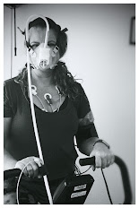
HOW DOES IT WORK: The biofeedback sensors "learn" about your specific physical signs and symptoms of stress and anxiety (ie. increased heart rate, body temperature, and muscle tension) through electrical sensors that are connected to specific areas of your body. During your session, your therapist will guide you through different mental exercises that may involve visualization, meditation, breathing, or relaxation techniques.  The ONDAMED Biofeedback System combines stimulation of gentle focused pulsed electromagnetic fields with Biofeedback. The feedback from the patient is done by monitoring the pulse rate and SpO2 displayed on the device. In addition, the practitioner palpates the patient’s radial pulse (aka vascular signal). This method allows personalizing stimulation of frequencies and sound along with placing applicators on most reactive stressed areas on the body for the patient’s fastest and lasting recovery.

"The ONDAMED device is an intelligent approach to providing stimulation with your body’s own communication language, which is electromagnetic. The brilliance of the ONDAMED method is the personalization of targeted localized frequency stimulation raising awareness to the patient to overcome stress and stress-related disorders."


"The ONDAMED device is an intelligent approach to providing stimulation with the body’s own communication language, which is electromagnetic. It’s not just going by symptoms or pathology or by what one knows about one frequency, but rather it is about combining a physical stimulation with biofeedback to personalize healthcare. Connecting with the patient by palpating the patient’s radial pulse while she/he receives ONDAMED’s therapeutic stimulation will give us the information as to what frequencies and where on the body these frequencies are needed most. It is overriding what we know or don’t know about the patient’s life health story. This method is comparable to a lie detector; we are accessing the individual’s innate wisdom by tapping into their communication pathways, and perhaps into part of their subconscious. Within minutes, the applicators can be placed on select areas for treatment. With biofeedback, we make the patient aware of what it is that they respond to—and where it is on the body that they need treatment. The patient’s autonomic nervous system is teaching the patient about their state of mind, their emotions, and biological consequences; this is all occurring while therapy is already running." 

- Dr. Silvia Binder


VIEWPOINTS

"As a PEMF medical protocol specialist, I’m experiencing the cellular response to PEMF with my patients every day. A dysfunction of the mitochondria can cause many disorders such as diabetes, Parkinson's disease and Alzheimer's. Research has shown that PEMFs recharge cells through ion exchange, bringing cellular energy back to its full potential and slowing the aging process of cells. Once cells are at their optimum potential, they are therefore able to operate more efficiently and for a longer period of time." - Joseph J. Toy / PEMF Medical Protocol Specialist.  (www.cliniquesneuroviesante.com)


NEW PROGRAM: Women's Diagnostic Network
Having access to the latest in compassionate experts and innovative diagnostic solutions for women is the first step to maintaining good health. Our alliance unites collaborative minds about the latest solutions from a wide range of modalities to offer optimum choices for all patients. Meet our current caregivers and educators for integrative care of women's chronic disorders. Together, we provide expansive research and info-sharing about all clinical options and protocols as well as testimonials and clinical viewpoints from active professionals in the field. 

The Women’s Diagnostic Network is a public informational resource backed by the Integrative Pain Healers' Alliance and the AngioFoundation. We are comprised of an all-volunteer collective of independent clinical experts, medical researchers and educators in specialized areas of clinical study. We support all areas of women's health interests including (but not limited to) CANCER CARE, ONCOLOGY, GYNECOLOGY, PHYSIATRY, DERMATOLOGY, NEUROLOGY, CARDIOLOGY & MENTAL HEALTH. Through clinical and academic collaboration, we unite to support improved patient evaluation, reporting on innovations and comprehensive diagnostic care. (visit WDN Webpage)

TOP HEADLINES
• 3/24 - Electromagnetic Breakthrough from Above our Northern Border: By Joseph J. Toy
• 3/16 - Essentials of SHOCKWAVE THERAPY: feat. Uran Berisha (IloveShockwave.com)
• 3/7 - Personalized Nutrition: Going beyond VitaminB12 for Vegan Diet Support: By: Dr. Bobbi Kline
• 3/7 - Integreative Medicine Review of Toxins
• 3/7 - The Sitting Culture: High Risk of Decline and Mortality: by Dr. Jonathan Kirschner
• 2/24 - Can PEMF Speed Up Healing Time? - by Dr. Jerry Dreessen
• 2/23 - Mental Health 101: Treating Incontinence Starts with Overcoming the STIGMA: by Dr. Bobbi Kline


REFERENCES:

1) Biofeedback, Mind-Body Medicine, and the Higher Limits of Human Nature:  https://www.aapb.org/i4a/pages/index.cfm?pageID=3383

2) https://www.ondamed.net/en/ondamed-biofeedback


Disclaimer & Copyright Notice: The materials provided on this website are copyrighted and the intellectual property of the publishers/producers (The NY Cancer Resource Alliance/IntermediaWorx inc., The AngioFoundation and Bard Diagnostic Research & Educational Programs). It is provided publicly strictly for informational purposes within non-commercial use and not for purposes of resale, distribution, public display or performance. Unless otherwise indicated on this web based page, sharing, re-posting, re-publishing of this work is strictly prohibited without due permission from the publishers.  Also, certain content may be licensed from third-parties. The licenses for some of this Content may contain additional terms. When such Content licenses contain additional terms, we will make these terms available to you on those pages (which his incorporated herein by reference).The publishers/producers of this site and its contents such as videos, graphics, text, and other materials published are not intended to be a substitute for professional medical advice, diagnosis, or treatment. For any questions you may have regarding a medical condition, please always seek the advice of your physician or a qualified health provider. Do not postpone or disregard any professional medical advice over something you may have seen or read on this website. If you think you may have a medical emergency, call your doctor or 9-1-1 immediately.  This website does not support, endorse or recommend any specific products, tests, physicians, procedures, treatment opinions or other information that may be mentioned on this site. Referencing any content or information seen or published in this website or shared by other visitors of this website is solely at your own risk. The publishers/producers of this Internet web site reserves the right, at its sole discretion, to modify, disable access to, or discontinue, temporarily or permanently, all or any part of this Internet web site or any information contained thereon without liability or notice to you.

Tuesday, February 15, 2022

A PEMF SUCCESS STORY: "GETTING MY LIFE BACK FROM CFS"

Interview by: Lennard Gettz, Ed.D & Roberta Kline, MD
Edited by: Dr. Robert L. Bard

Introduction

In support of personal leadership and proactive health, The Integrative Pain Healers Alliance applauds Ms. Suzanne Wheeler of Minneapolis, Minnesota as our Researcher of the Month.  After years of suffering a life-altering disorder that currently continues to challenge the scientific community of its root causes, Suzanne explored “outside the conventional box” of opioid prescriptions until she uncovered the one remarkable solution that got her back on her feet and joining life again.  Invoking CHANGE against all odds by diligently searching for what’s beyond the convenient takes courage and conviction.  It is this level of leadership that defines Alternative Health and Wellness to educate others about new answers that offer better results.

SHOT DOWN BY CFS
In 2017, I was diagnosed with Myalgic Encephalitis or Chronic Fatigue Syndrome. I had been essentially home bound for about two years and probably bed bound about 50% of my time. CFS was found to be the source of my 'all over body' pain- and so started my road to seeking out more targeted treatments. 

Life before CFS was very active. I worked in corporate America for 17 years as a regional manager for a large building facilities company and managed a team of about 3000 people. Prior to that, I was in the United States army for a decade. I was a Blackhawk pilot and a C 12 airplane pilot in the military before I entered corporate America. 

I started to feel significant pain after the birth of my 3rd daughter in Oct 2002.  I had a traumatic delivery due to a condition called placenta previa.  I lost over half my blood volume during the birth and received extensive blood transfusions.  I never felt the same after that experience.  About a week after delivery, the pain throughout my body started.  Over the years – it became worse and worse, and the fatigue became debilitating.  I kept pushing through with my job and family responsibilities which may have damaged my body even further.  


CFS is not an easy problem to resolve. I spent extensive time at Stanford University's chronic fatigue clinic with some of their top specialists.  I was also sent to the Workwell Foundation (CA) where I underwent a two day CPET (image insert) a cardiopulmonary exercise testing where they were able to determine what my anaerobic threshold was my VO2 max.  It was there that the clinicians found the extent of my metabolic dysfunction; my reports showed a VO2 reading equivalent to that of about a 95 year old. 

I had the kind of pain that radiated all over- throughout my spine, throughout my hips, legs, my knees, ankles, feet also throughout my arms. I had, peripheral neuropathy in my hands. I had trouble moving my hands and trouble typing and then also trouble walking. It developed to the point where in 2015 I was using a cane in my late forties to walk. I was desperate to get pain relief for more than a decade. I also had compression in my lower spine and my L4, L5, and also in my thoracic region (T 11 & 12) that caused extensive pain.  It wasn't an easy thing to figure out but through MRI, you can see the physical deformities- where imaging helped make everything pretty clear. But as to why it radiated through my body like it did, it took a lot of years to find out. 

DESPERATE FOR PAIN RELIEF
Since 2002, I tried everything- modalities from opioids to hip injections to spinal injections from a pain clinic. I saw chiropractors, I had acupuncture, tens units, electrical stim, laser treatments, extensive cranial psychotherapy, massage, a lot of ice and heat, a massage chair and a hot water Jacuzzi (which was the one thing that I responded to the best).

All pain relief from this point was temporary.  The base pain would eventually return and continue to get progressively worse. Years into this, it didn't seem like there was a lot left for me to try.  I learned that my condition was based on a specific virus that destroys your metabolic system and your body doesn't have the ability to oxygenate for energy. I was extremely tired all the time with post exertional malaise and my anaerobic thresholds became very low.  Our body builds up lactic acid with any exertion, with even minimal amounts of exertion. 

TAKING CHARGE: RESEARCH OUTSIDE THE BOX
In 2017, I learned about PEMF and its concepts of getting to the cellular level by helping my cells oxygenate.  Reading the many testimonials about it, it seemed to make better sense than the other modalities I tried so far.  User stories matched up with my symptoms and what I had been told by doctors was happening to my body. My sister introduced me to the idea upon seeing a presentation at a horse event, believe it or not. I became intrigued and acquired a rental and eventually purchased my  own model.

ALTERNATIVE HEALTH
I have HAD some wonderful physicians who are experts in their lane. At the height of my illness, I was seeing six different doctors who had me on so many medications (10 or 12 prescriptions) and I was still bed ridden- hence, nothing was working. 

Before I started using the PEMF, I had been essentially home bound for about two years and probably bed bound about 50% of my time. And if I was not in bed, I was seeking pain relief, usually in a hot bath. I would say within two weeks, the pain throughout my body had significantly subsided. I still had a little bit of joint pain but the overall soft muscle pain was GONE.  I had been taking opioids for over a decade prior, and I practically eliminated all that. It was, it was pretty unbelievable. Pain was a huge factor.  I wasn’t changing my doctors - but the one thing we all agreed on is that they found sudden improvement in me.  In my gut, I was confident of the changes were from the use of PEMF.


SUZANNE TODAY
I live a completely different life. To be clear, I'm not healed. I still have to live a more gentle life- certainly nothing like I used to do back in my army days or my early years working in corporate America- running and teaching aerobics and doing things like that. I still am not there to be able to do all that. The disease is still there, but the symptoms are managed and I have eliminated all the harsh, addictive medications. I am definitely getting so much more out of life without pain pills as opposed to being bed ridden and walking with a cane.

For anyone suffering like this, it's worth looking into. You want the expertise from the medical world, but you should blend the holistic treatments that are available- and then research it. If you search PEMF- read up on it! You'll find so many clinical studies and user testimonials that are coming up on that for all different ailments (not just mine) and they're seeing results. 

Thanks to PEMF, I'm able to participate with my family. I'm able to travel, go to the beach and take wonderful walks again. My sisters and I started my own business helping horses and people (with PEMF) be more pain-free and traveling again. It's hard not to be passionate about something that has that effect on your life. My husband says he got his wife back.


SPECIAL THANKS
The editors of this spotlight proudly gives thanks to Ms. Suzanne Wheeler for her generosity in sharing her story and her resources with us.  Additional thanks to Dr. Jerry Dreessen of the AOPP (Association of PEMF Professionals) and Pat Ziemer of Magnawave Inc. and Aura Wellness PEMF for coordinating our interviews, shared countless materials and conducted unending support to help our educational program bring new light to PEMF technology for chronic disorders and supportive testimonials in alternative therapeutics.   


ScienceNews Extra
Commentary on CFS/ME (Chronic Fatigue Syndrome and Myalgic Encephalomyelitis
By: Dr. Bobbi Kline

One of the key underlying findings in conditions causing chronic mental or physical fatigue is dysfunction of the mitochondria. Mitochondria are small structures located in the nucleus of every cell, and every cell contains thousands of them. These powerhouses produce the energy our bodies need to carry out every function in the form of ATP (adenosine triphosphate). When mitochondria don’t function at their best, or too many of them are destroyed, our energy levels suffer. While this can be due to inherited genetic disorders, most often it is seen as the result of chronic damage over time.

Many of the biochemical reactions in our bodies produce toxic versions of oxygen, hydrogen and nitrogen, including how we make ATP. These toxic molecules, which we call free radicals, have to be neutralized so they don’t damage the mitochondria. Our bodies have powerful antioxidant defenses to keep these in check. But when these protective systems become overwhelmed by too many free radicals, oxidative stress results and mitochondria are damaged.


IN THE NEWS

THE 2022 POST-MILITARY CRUSADERS COALITION 
2/11/2022- A 2022 initiative by community leaders launched the PMCC or Post Military Crusaders Coalition to launch an action plan for health resources for injured American veterans. Similar to the First Responders Cancer Resource project, this campaign supports all veteran advocates and service members support organizations by offering educational initiatives, alternative therapeutic modalities, sustainable diagnostic technologies and clinical research programs. 


CONTRIBUTORS

BOBBI KLINE, MD 
(Educational Dir. /Women's Diagnostic Group)
Dr. Kline is a board-certified ObGyn physician, Integrative Personalized Medicine expert, consultant, author, and educator whose mission is to change how we approach health and deliver healthcare. She helped to create the Integrative & Functional Medicine program for a family practice residency, has consulted with Sodexo to implement the first personalized nutrition menu for healthcare facilities, and serves as Education Director for several organizations including the Women’s Diagnostic Health Network, Mommies on a Mission. Learn more at https://bobbiklinemd.com 

 
ROBERT L. BARD, MD  (Diagnostic Imaging Specialist)
Having paved the way for the study of various cancers both clinically and academically, Dr. Robert Bard co-founded the 9/11 CancerScan program to bring additional diagnostic support to all first responders from Ground Zero. His main practice in midtown, NYC (Bard Diagnostic Imaging- www.CancerScan.com) uses the latest in digital Imaging technology has been also used to help guide biopsies and in many cases, even replicate much of the same reports of a clinical invasive biopsy. His most recent program is dedicated to the reporting of mental health diagnostic and innovative solutions including the use of modern neuromagnetic technologies and protocols in his MEDTECH REVIEWS program. 


Our friends and supporters:


Disclaimer: The information (including, but not limited to text, graphics, images and other material) contained in this article is for informational purposes only. No material on this site is intended to be a substitute for professional medical advice or scientific claims. Furthermore, any/all contributors (both medical and non-medical) featured in this article are presenting only ANECDOTAL findings pertaining to the effects and performance of the products/technologies being reviewed - and are not offering clinical data or medical recommendations in any way. Always seek the advice of your physician or other qualified health care provider with any questions you may have regarding a medical condition or treatment and before undertaking a new health care regimen, never disregard professional medical advice or delay in seeking it because of something you read on this page, article, blog or website.

Monday, February 7, 2022

PEDIATRIC MYOCARDITIS & NATIONAL DETECTION MOVEMENT (Part 1)

Written by: Dr. Robert L. Bard

Since the advent of Covid-19 Long Haul studies in 2021, the medical diagnostic community shifted into overdrive- seeking out all available screening and examination protocols to assess health problems called POST-ACUTE SEQUELAE (PASC). One of the recent Covid-19 related headliners is the rise in cases of MYOCARDITIS in children 16 years and under. CDC Reports link the pathological impact with covid infection since it is proven that Viral infections are a common cause of myocarditis. 

Between early 2020–2021, patients with Covid-19 had nearly 16 times the risk for myocarditis[1]. According to the CDC, in a study of myocarditis cases, 2,116 (41.7%) had a history of Covid-19.  In addition, cases of myocarditis reported to the Vaccine Adverse Event Reporting System (VAERS) indicated links between Myocarditis and Pericarditis to come from the mRNA Covid-19 vaccination (especially in male adolescents and young adults) more often after the second dose.[2].

Myocarditis is defined as an inflammatory disorder of the heart muscle (myocardium) leading to cardiac dysfunction. It is also recognized as myocardial cell death [3]. Checkups for this also reviews for PERICARDITIS (the inflammation of the outer lining of the heart). Various causes of myocarditis includes: Viral Infectious including adenoviruses, echoviruses, enteroviruses like the coxsackie viruses. In addition, predisposition can occur from those with Autoimmune diseases such as Celiac disease, Churg-Strauss syndrome, Crohn disease, Kawasaki disease, lupus, rheumatoid arthritis, sarcoidosis etc. (See NIH chart for full list of causes- [4])


STANDARDIZED DIAGNOSTICS

As with any critical disorder, detecting early stages of myocarditis allows for a higher opportunity to treat and even eliminate the health risk.  In children, symptoms include: Fever, Fainting, Breathing difficulties, Rapid breathing, Chest pain and Rapid or irregular heart rhythms [5]. In adults, symptoms range from chest pain, shortness of breath, at rest or during activity and fluid buildup with swelling of the legs, ankles and feet. To prevent possible heart damage, a cardiologist may order one of a number of imaging options:

▪ Electrocardiogram (ECG or EKG)
▪ Chest X-Ray
▪ Heart MRI
▪ Blood Tests
▪ Doppler Ultrasound for Acute Myocarditis
▪ Cardiovascular MR Elastography (MRE)
▪ Ultrasound Elastography

See expanded details on diagnostic protocols, visit:  http://pediatricscan.com/myocarditis.html


MedNews Extra
Saving Lives Through Advocacy & Research:
FOR ELYSA FOUNDATION

Around the first week of Feb, 2022, our clinical diagnostic researcher, Dr. Robert Bard launched his PediatricScan.com 2.0 in NYC‐ which included a Pilot program for Myocarditis Screening through the use of advanced Doppler Ultrasound Imaging.  To establish the clinical network for this program is to connect with ICU specialists & Cardiologists as well as all associations supporting Myocarditis research.

We met the directors of a remarkable national advocacy foundation called FOR ELYSA FOUNDATION‐ a non‐profit organization dedicated to promoting Education, providing Light, and supporting Research in the areas of viral myocarditis and pediatric sudden cardiac arrest. (www.ForElysa.org). Mrs. Jana Rojas and husband Jaime Rojas from Kansas City developed this organization inspired by the loss of her vibrant little girl,  Elysa Louise Rojas who passed away at the tender age of two years old. " In Elysa’s case, a common childhood virus was responsible for her myocarditis. The virus either attacked Elysa’s heart directly or caused her immune system to attack her heart muscle in a “friendly fire” fashion while trying to fight the virus. The inflammation in her heart increased drastically and very quickly to the point of sudden cardiac arrest. Doctors and scientists do not fully understand the mechanisms within the body that cause a virus to “go haywire” in the immune systems of individuals with myocarditis. There is currently no way to predict when/if this will occur."


The FOR ELYSA FOUNDATION is one of our first advocacy friends in pursuit of bringing national awareness and supportive clinical research for myocarditis diagnostics and prevention. According to the ForeElysa.org website, Myocarditis is a disease marked by inflammation and damage of the heart muscle. There are many causes of myocarditis, including viral infections, autoimmune diseases, environmental toxins, and adverse reactions to medications. The most common cause of myocarditis in North America is viral infections. Myocarditis usually attacks otherwise healthy people. It is believed that 5 to 20% of all cases of sudden death in young adults are due to myocarditis. Although the exact incidence of myocarditis is not known, it is estimated that approximately 343,000 people die of myocarditis and its major complication, cardiomyopathy, each year. The prognosis is variable but chronic heart failure is the major long term complication. Myocarditis and the associated disorder of idiopathic dilated cardiomyopathy are the cause of approximately 45% of heart transplants in the United States.  

Materials in this excerpt are published with express consent from The For Elysa Foundation.  For complete Information, visit www.FORELYSA.org


HOW & WHEN TO CHECK FOR PEDIATRIC MYOCARDITIS
By: Jana Rojas
The tricky thing with myocarditis being virally mediated is that Elysa could have had a heart scan a week before she died (the day before she contracted the virus that wrecked her heart), and it would have been normal. I am hesitant to insinuate that imaging could "clear" a patient and provide a clean bill of health without noting that this can and does occur spontaneously after viral infections, and so testing while ill or post-virally is actually the key message and window of opportunity for myocarditis detection.
In my mind, the primary role for cardiac diagnostic imaging as it related to myocarditis specifically would be for: 
1) acutely ill children in ED/urgent care/hospital inpatient settings
2) children exhibiting the signs and symptoms you have outlined (fainting, sudden fatigue, shortness of breath, chest pain, palpitations), 
3) after known Covid or other viral infection with prolonged or delayed healing (ie ongoing fatigue, shortness of breath, etc)  
4) and possibly PRE-PARTICIPATION SPORTS PHYSICALS. The pre participation screenings would be enhanced cardiac screenings in general to ideally pick up congenital heart defects and other concerns as well as myocarditis. 


TROUBLESHOOTING MYOCARDITIS
By Bobbi Kline, MD (Integrative Physician / Genomic Research Specialist)

As a mom, my heart grieves for parents, including Elysa’s, who suffer such devastating tragedy. It’s the worst thing you hope never happens to your child, and I truly admire parents who turn a tragedy into something positive. It requires such amazing strength, courage and grace. As a physician, I find my self immediately asking "Why do these things happen? How can we predict or prevent them?" As clinicians, we look for patterns to help guide diagnosis and treatment. We know what to expect, but sometimes they can lull us into a false sense of security. Childhood viruses, as any parent knows, are an expected part of those early years. 
But what happens when they turn out to be something more? That’s where pattern recognition is crucial. When something obviously falls outside those patterns, it’s a signal to question and go deeper. But what happens when you don’t even recognize that deviation? What if something is so uncommon or so subtle that it’s hard to detect among all the noise? Post-viral myocarditis is one of those conditions, and I’m glad to see it now in the spotlight. Raising awareness is a key first step. While COVID-19 has certainly helped to highlight this condition, it goes further than COVID. Many common childhood viruses have been implicated in causing myocarditis, but most people are completely unaware. I admit that it was not something I ever really thought about as my kids were growing up. And I am not alone. Educating clinicians as well as parents on what to look for, when to be alarmed, when to go deeper is crucial. This alone will save lives. 


But it’s only the first step. We also need better tests and tools to quickly and easily identify who is at risk, and better treatments for helping these children. This requires a multidisciplinary approach that includes better diagnostics including noninvasive technology, along with effective medications and other treatments. It also includes the burgeoning field of genomics and personalized medicine, both to provide a better understanding of the why, as well as a powerful tool to predict and prevent. For, at the heart of this, is understanding each child’s uniqueness in a way that empowers.  Two studies have been published this year that have the potential to leverage the power of DNA to identify who is at risk for developing myocarditis after a viral infection. Not only that, but also which of those children are most likely to recover, and therefore need fewer interventions, and which of those children are most at risk for sudden death and require much more intensive treatment and support. And, in today’s world, we also need the power of legislation to make sure everyone has access to this higher level of care. There is much promise to change the trajectory of this devastating illness, and it is only through advocacy such as this that it will happen. 


1) Morbidity and Mortality Weekly Report (MMWR): Association Between Covid-19 and Myocarditis Using Hospital-based Admin Data 3/2020-1/2021) https://www.cdc.gov/mmwr/volumes/70/wr/mm7035e5.htm
3) MR Imaging of Myocardial Infarction | RSNA-Radiological Society of North America  / https://pubs.rsna.org/doi/10.1148/rg.335125722
4) The Diagnostic and Clinical Approach to Pediatric Myocarditis: A Review of the Current Literature (NCBI/NIH)  https://www.ncbi.nlm.nih.gov/pmc/articles/PMC6352488/
5) Diagnosis and Management of Myocarditis in Children (American Coll. of Cardiology) https://www.acc.org/latest-in-cardiology/ten-points-to-remember/2021/07/09/17/31/diagnosis-and-management-of-myocarditis

Disclaimer: The information (including, but not limited to text, graphics, images and other material) contained in this article is for informational purposes only. No material on this site is intended to be a substitute for professional medical advice or scientific claims. Furthermore, any/all contributors (both medical and non-medical) featured in this article are presenting only ANECDOTAL findings pertaining to the effects and performance of the products/technologies being reviewed - and are not offering clinical data or medical recommendations in any way. Always seek the advice of your physician or other qualified health care provider with any questions you may have regarding a medical condition or treatment and before undertaking a new health care regimen, never disregard professional medical advice or delay in seeking it because of something you read on this page, article, blog or website. None of the information provided should be interpreted to be or is meant to be medical advice, suggestions, or counseling.

Thursday, January 20, 2022

INHERITING CANCER (feat: Genetic & Genomic Testing)

CANCER HEREDITY
(Originally published 11/2020) Cancer is a genetic disease—that is, cancer is caused by certain changes to genes that control the way our cells function, especially how they grow and divide. Genes carry the instructions to make proteins, which do much of the work in our cells. Certain gene changes can cause cells to evade normal growth controls and become cancer. For example, some cancer-causing gene changes increase production of a protein that makes cells grow. Others result in the production of a misshapen, and therefore nonfunctional, form of a protein that normally repairs cellular damage.  

Inherited genetic mutations play a major role in about 5 to 10 percent of all cancers. Researchers have associated mutations in specific genes with more than 50 hereditary cancer syndromes, which are disorders that may predispose individuals to developing certain cancers. Genetic tests for hereditary cancer syndromes can tell whether a person from a family that shows signs of such a syndrome has one of these mutations. These tests can also show whether family members without obvious disease have inherited the same mutation as a family member who carries a cancer-associated mutation. [1]


Everyone has two copies of each set of genes from both parents. If a person inherits a mutation in a Lynch syndrome gene, they still have the normal copy of the gene from the other parent. Cancer occurs when a second mutation affects the normal working copy of the gene, so that the person no longer has a copy of the gene that works properly. Unlike the inherited Lynch syndrome mutation, the second mutation would not be present throughout the person’s body, but would only be present in the cancer tissue. However, not everyone with Lynch syndrome will get cancer. You and your family members are more likely to have Lynch syndrome if your family has a strong history of colorectal cancer. Family members who inherit Lynch syndrome usually share the same mutation. If one of your family members has a known Lynch syndrome gene mutation, other family members who get genetic testing should be checked for that mutation. (source: CDC.gov)

GENETICS & GENOMICS: SIMILAR BUT DIFFERENT

Excerpted from https://robertaklinemd.com


Genetics and genomics sound alike and are often used interchangeably. While they both deal with changes in our DNA, there are important scientific and clinical distinctions.

GENETICS CAME FIRST
The science of genetics is classically defined as the study of heredity, or how traits are passed on from one generation to the next. While modern day genetics evolved from Gregor Mendel’s pea experiments in the mid-1800s, it wasn’t until James Watson and Francis Crick discovered the actual structure of DNA in 1953 that we began to understand exactly how it works. 

DNA contains the genetic code, the blueprint for everything that happens in our bodies. DNA is short for deoxyribonucleic acid, and it is composed of long strands made up of four different nucleotides called adenine, guanine, thymine, and cytosine, or A, G, T, and C for short. It exists as two long strands that are paired in a specific manner. The DNA is grouped into functional units called genes, which can comprise either a small or large section of DNA.

This DNA code is incredibly complex, and in order to contain all this information, the amount of DNA we each have in our cells is tremendous. In fact, every cell contains about 6 feet of DNA. That’s enough DNA in one person that, if stretched out, it would be long enough to go to the sun and back more than 300 times… or go around the earth’s equator 2.5 million times!

That’s where chromosomes come in. The DNA is compressed into tightly coiled structures called chromosomes in order to fit into our cells. We each have 23 pairs of chromosomes, and they are stored in a special compartment of the cell called the nucleus. 

While that solves the storage problem, it creates a different problem – how to access the instructions encoded in the DNA. Every time that cell needs to replicate itself to replace dead cells in an organ or tissue, or we need to make a certain protein, portions of the chromosome need to be unwound to expose the needed genetic code. But sometimes during that process mistakes are made, and the change in the DNA can be mild or catastrophic.

GENETICS: BLACK & WHITE
Genetics traditionally deals with changes in the DNA sequence called mutations that are inherited in a predictable manner. These can involve small sections of DNA all the way up to whole chromosomes. Medical genetics has traditionally focused on health conditions that are due to inherited mutations in single genes (Huntington disease), in whole chromosomes (trisomy 21 or Down syndrome) or are associated with birth defects or developmental disabilities. 

With a traditional, single-gene disorder such as Tay-Sachs disease, genetic information is obtained from a patient as well as his or her immediate family members and relatives. The information is then used to diagnosis or predict the risk of an inherited genetic disorder passing from one generation to another. For many single-gene disorders, there are very limited medical interventions available. Genetic disorders are individually rare, and they account for about 5% of all human disease.

That meant 95% of the diseases were due to some other mechanism. In an effort to better understand how DNA contributes to health, scientists realized that genetic mutations were only part of the story. The Human Genome Project was conceived in the late 1980’s to provide answers.

One of the biggest take-aways from the Human Genome Project was that humans share 99.5% of their genome. But while this means we are far more alike with all the genes we have in common, that 0.5% difference is significant. The small differences are what make us each unique.


Genomic Medicine: It is now possible to use results from clinically-based genomic testing to evaluate a person’s disease susceptibility, and develop evidence-based, personalized intervention strategies to reduce those risks. These strategies include DNA-directed lifestyle modifications, dietary recommendations, nutritional supplements and/or exercise, all of which influence how these genes function to create health or disease. Biomarker testing can then be used to evaluate whether the intervention is efficacious. With this approach, the guess-work and inefficiencies of trial-and-error strategies are greatly reduced, leading to better health more quickly and cost-effectively.

A comprehensive test can provide gender-specific reports to create personalized programs for many health conditions, including:

  • Prevent ER+ breast cancer or its recurrence in women diagnosed with the condition
  • Reverse osteopenia and osteoporosis
  • Prevent or treat Type II diabetes, heart disease, stroke, metabolic syndrome and obesity.
  • Optimize athletic performance
  • Customize nutrient requirements and resolve nutritional paradoxes
  • Prevent, treat and manage depression and anxiety
  • Plus many more health and wellness issues
  • Genomic Medicine is personalizing healthcare with precision. 

To see complete article, visit: https://robertaklinemd.com/


About the Author

BOBBI KLINE, MD 
(Educational Dir. /Women's Diagnostic Group)
Dr. Kline is a board-certified ObGyn physician, Integrative Personalized Medicine expert, consultant, author, and educator whose mission is to change how we approach health and deliver healthcare. She helped to create the Integrative & Functional Medicine program for a family practice residency, has consulted with Sodexo to implement the first personalized nutrition menu for healthcare facilities, and serves as Education Director for several organizations including the Women’s Diagnostic Health Network, Mommies on a Mission. Learn more at https://bobbiklinemd.com 


Directory of Inherited Cancer Syndromes source: NIH LINK
• BRCA1 / BRCA2 - breast & ovarian cancer
• Cowden Syndrome
• Diaphyseal medullary stenosis
• Duodenal carcinoid syndrome
• Endolymphatic sac tumor
• Familial adenomatous polyposis
• Familial isolated pituitary adenoma
• Gardner syndrome
• Hereditary diffuse gastric cancer
• Hirschsprung disease ganglioneuroblastoma
• Langerhans cell histiocytosis
• Li-Fraumeni syndrome
• Lynch syndrome
• Multiple endocrine neoplasia:
- (type 1
 | type 2A | type 2B)
• MYH-associated polyposis
• Oslam syndrome
• Paraneoplastic Neurologic Disorders
• Perlman syndrome
• Pheochromocytoma | islet cell tumor synd.
• Premature aging Okamoto type
• Stewart Treves syndrome
• Von Hippel-Lindau disease
• WAGR syndrome

 LYNCH SYNDROME is a hereditary cancer condition in which a mismatched repair gene, which ordinarily repairs errors in DNA duplication, is defective. As a result, individuals are predisposed at a very high lifetime risk of cancer, including an up to 85% risk for colorectal cancer, 65% risk for uterine cancer, 19% risk for gastric cancer, 13% risk for ovarian cancer and a higher than average risk for other cancer including cancers of the liver, gallbladder, kidney, bladder, prostate, pancreas, skin, brain and breasts. With genetic testing, there is hope...once diagnosed, annual cancer screenings take place and cancers can be treated or removed before becoming life threatening. By knowing our family history and having a great medical team, we live longer than ever before...as long as anyone else!


An Introduction to Lynch Syndrome
By: Lindy Bruzzone

I was diagnosed in 2007 with colorectal cancer which was tied to my having Lynch Syndrome.  There are tens of thousands of people now diagnosed with this genetic disorder which means this is NOT a RARE disorder like most might think.  It came from a defective mismatch repair gene, where instead of repairing errors in DNA replication, it puts in different proteins and create more errors than it tries to keep doing. 

I produced this video 10 years after I lost lost my father. It was just so difficult for me to lose him that way. The day before he died, he said to me, "the doctors think it's hereditary, go get checked and tell your brother and sister, to get checked."  After his funeral, my brother and I started looking for the right cancer center to get help. Knowing my father's reports, my surgeon and other doctors jumped in to get me tested, and sure enough--- it was positive. 

That's why Lynch Syndrome International was developed because nobody was getting diagnosed. NOW they are! And then when Color Genomics was developed (a Steve Jobs startup offering for FREE genetic testing), suddenly everybody started getting tested because they could finally afford it. They didn't want insurance companies involved. It took care of every barrier that we have to testing for hereditary cancers. 

tbc


References

1) Cancer Heredity / NIH: https://www.cancer.gov/about-cancer/causes-prevention/genetics

Thursday, December 16, 2021

MEDTECH REVIEW: Fluorescence Imaging for Inflammation and Infection (official)

Written by: Dr. Robert L. Bard

Upon review of all the imaging technologies available, clinicians are constantly in search for what provides us the most accurate quantitative data from the standpoint of non‐invasive safety. In the case of diagnosing cancers and bacterial infections, we are now exploring the potential of the intelligence behind bioluminescence and fluorescence (or autofluorescence) probes.

From a 2019 study (by Raizman et al) about use of this technology (MolecuLight i:X) with debridement, scans of 22 wounds were conducted under standard and violet light in patients after debridement. Scans, performed non-invasively at the point-of-care, demonstrated remaining bacteria/biofilm signal in 100% of wounds after sharp debridement, triggering additional removal of tissue. [1] 

Re-scanning demonstrated a marked decrease or complete removal of bacteria in most wounds, yet a subset showed persistent or increased bacterial signals post debridement. The authors, aided by the knowledge gained from this technology,  proposed “the subgroup with persistent bacterial fluorescence post-debridement was at increased risk of deep compartment infection and required more frequent debridement and/or antibiotics.” [1]. See example of this in figure 2 (tissue in green, bacteria in red). Others have demonstrated ability to markedly decrease antibiotic usage using this technology, showing great potential for stewardship efforts [2].

Currently, the technology can take direct images of the skin or mucosal surface to indicate bacteria on the tongue or oral lining. Exploratory reviews of this imaging device also shows possible benefits in scanning potential biopsy material with violet light at the tissue and see if it's suggestive for cancer or inflammatory disease. 


SCAN 1: The Test Drive

Autofluorescence has been proven and used worldwide to show inflammatory changes and bacterial infection. It's used to find both Gram‐negative bacteria and Gram‐positive bacteria, aerobic and anaerobic. (Fig‐1) The illustrated scan shows the normal skinfolds as green and the skin image is homogeneous (there's nothing dark). (Fig‐2) The scan of the finger on the opposite hand which the patient expressed chronic irritation. Upon initial observationit appears that Autofluorescence may detect inflammation through the upper dermis, a change documented visually and by ultrasound imaging as minimal epidermal thickening associated with inflammatory skin disease (mild type). When we turned on the fluorescence scan, the exact one centimeter area of redness on the skin corresponded to the one centimeter darkened area on the finger.


SCAN 2: CLINICAL IMAGING & OBJECTIVE DATA MEASURING WITH ULTRASOUND 
(Fig-3) For my diagnostic research projects in dermal lesions and infections, one of my "weapons of choice" is the doppler ultrasound.  From tumors to traumas, radiologists and clinicians rely on feature-rich computerized systems that provided remarkable patient data for its non‐invasive and real-time view of inflammatory disease to align with our study of the Autofluorescence for its work on atopic dermatitis and other viral diseases (ie. Covid-19) which may affect the skin and be sampled with this noninvasive technology. Since it hasn't been clinically documented in the dermal tissues, this is an opportunity to form a new type of imaging that is non-invasive to consider avoiding biopsies on children.

In the case of Fig-3, I used a high-frequency setting on a popular handheld point of care ultrasound probe to recognize a 0.3cm depth scan of the same area of the finger that was previously scanned by the fluorescence device. The top right arrow (1a) shows the enlarging dark stripe indicating inflammatory thickening or the epidermis. The middle grey area (arrow 1b) represents subdermal inflammation of subcutaneous tissue or mild inflammatory skin disease -  confirming the prior evaluation.

EXPANSION POTENTIAL: RESEARCH 
Fluorescence imaging is proven to have a four‐millimeter depth of diagnostic accuracy in the tongue or mucous membranes‐ similar to the cervix or the vagina or the intestine lining. There is a rich literature of its use in skin wounds and wound infection. However, it's use in skin diseases has not been fully explored‐ so this is a beta test to see if and how it will work in non‐invasive diagnosis of inflammatory skin disease, especially in children. Exploring this technology is of great importance because dermatitis is recognized as a major pediatric disorder. Also disabling inflammatory chronic skin disease (psoriasis, rosacea) are a major focus of successful treatment by pharmacology companies.


AUTOFLUORESCENCE FIELD TEST   
 
by: Randall Weisel, DDS (Excerpt from Journal of Dentistry and Oral Sciences/ "Auto Fluorescence Allows Us to Detect Early Signs of Oral Cancer and Much More")

Review of autofluorescence enables us to objectively examine the oral cavity for inflammation and infection. Many systemic diseases are perpetuated by microorganisms that colonize in the oral environment. They enter the cardiovascular system by enzymatic processes that open the oral mucosa to allow their entry. A majority of the microbes are anaerobes and/or facultative anaerobes. When they enter the host, they metabolize blood. Their waste by products contains iron elements within a compound called porphyrin. Porphyrin will fluoresce when exposed to certain wavelengths of light. Healthcare providers can utilize this natural occurring process to objectively see these harmful pathogens. This may indicate that the host has a Sleep Related Breathing Disorder (SRBD). Sleep apnea is a primary disorder of SRBD’s. This technology offers medical and dental fields a screening tool for a pandemic healthcare problem.

Pathogenic microbial presences and the size of the colony (the bioload) can be relatively determined [19]. Fluorescence visualization of red-orange means bacterial pathogens are present. Presence of bluish green/bright white indicates presence of Pseudomonas [19]. The conditions for these pathogens to exist can occur within individuals with Sleep Related Breathing Disorders (SRBD). Sleep Apnea is the most prevalent of these disorders [20]. Researchers should focus their attention to the dorsum of the tongue ecosystem. Autofluorescence, performed with technology that utilizes electromagnetic spectral ranges and special filters that are components of the Velscope, allows visualization of pathogenic microbes. Scientific research demonstrates that oral pathogens (Pg, Aa, and Fn) may be direct and/or indirect causative agents of systemic diseases. These oral pathogens cause inflammation and infection.


SPECIAL THANKS to Dr. Monique Rennie, Director of Scientific Affairs and Global Engagement at Moleculight for her generous technical support and assistance greatly added  to the quality and integrity of this feature article. 


REFERENCES:

1) Raizman R et al. Use of a bacterial fluorescence imaging device: wound measurement, bacterial detection and targeted debridement. J Wound Care (2019). https://pubmed.ncbi.nlm.nih.gov/31825778/

2) Price N. Routine fluorescence imaging to detect wound bacteria reduces antibiotic use and antimicrobial dressing expenditure while improving healing rates: retrospective analysis of 229 foot ulcers. Diagnostics 2020;10:927.

19) Le, L.; Baer, M.; Briggs, P.; Bullock, N.; Cole, W.; DiMarco, D.; Hamil, R.; Harrell, K.; Kasper, M.; Li, W.; et al. Diagnostic Accuracy of Point-of-Care Fluorescence Imaging for the Detection of Bacterial Burden in Wounds: Results from the 350-Patient Fluorescence Imaging Assessment and Guidance Trial. Advances in Wound Care. 2021. https://pubmed.ncbi.nlm.nih.gov/32870774/

20) Sleep-Disordered Breathing.chapter 23.Thoracic.org. 237-247. Boillot A, Demmer RT, Mallat Z, Sacco RL, Jacobs DR, Benessiano J, et al. Periodontal microbiota and phospholipases: the oral infections and vascular disease epidemiology study (INVEST). Atherosclerosis. 2015;242(2):418-23. PubMed | CrossRef

21) Bale BF, Doneen AL, Vigerust DJ. High-risk periodontal pathogens contribute to the pathogenesis of atherosclerosis. Postgrad Med J. 2017;93(1098):215-20. PubMed | CrossRef

22. Boillot A, Demmer RT, Mallat Z, Sacco RL, Jacobs DR, Benessiano J, et al. Periodontal microbiota and phospholipases: the oral infections and vascular disease epidemiology study (INVEST). Atherosclerosis. 2015;242(2):418-23. PubMed | CrossRef

23) Desvarieux M, Demmer RT, Jacobs Jr DR, Rundek T, Boden-Albala B, Sacco RL, et al. Periodontal bacteria and hypertension: the oral infections and vascular disease epidemiology study (INVEST). J Hypertens. 2010;28(7):1413. PubMed | CrossRef


Disclaimer & Copyright Notice: This article/website does not provide medical advice. The information, including, but not limited to text, graphics, images and other material contained on this blogsite are for informational purposes only. No material on this site is intended to be a substitute for professional medical advice, diagnosis or treatment. Furthermore, any/all contributors mentioned in this article are presenting only ANECDOTAL findings of the effects of the products/technologies they are featuring- and are not offering clinical data or medical recommendations in any way. Always seek the advice of your physician or other qualified health care provider with any questions you may have regarding a medical condition or treatment and before undertaking a new health care regimen, never disregard professional medical advice or delay in seeking it because of something you read on this page, article, blog or website.

The materials provided on this website/web-based article are copyrighted 2021 and the intellectual property of the publishers/producers (The NY Cancer Resource Alliance/IntermediaWorx inc. and Bard Diagnostic Research & Educational Programs). It is provided publicly strictly for informational purposes within non-commercial use and not for purposes of resale, distribution, public display or performance. Unless otherwise indicated on this web based page, sharing, re-posting, re-publishing of this work is strictly prohibited without due permission from the publishers.  Also, certain content may be licensed from third-parties. The licenses for some of this Content may contain additional terms. When such Content licenses contain additional terms, we will make these terms available to you on those pages (which his incorporated herein by reference).The publishers/producers of this site and its contents such as videos, graphics, text, and other materials published are not intended to be a substitute for professional medical advice, diagnosis, or treatment. For any questions you may have regarding a medical condition, please always seek the advice of your physician or a qualified health provider. Do not postpone or disregard any professional medical advice over something you may have seen or read on this website. If you think you may have a medical emergency, call your doctor or 9-1-1 immediately.  This website does not support, endorse or recommend any specific products, tests, physicians, procedures, treatment opinions or other information that may be mentioned on this site. Referencing any content or information seen or published in this website or shared by other visitors of this website is solely at your own risk. The publishers/producers of this Internet web site reserves the right, at its sole discretion, to modify, disable access to, or discontinue, temporarily or permanently, all or any part of this Internet web site or any information contained thereon without liability or notice to you.