Sunday, April 17, 2022

2022 MedTech Review Update: TRANSCRANIAL DOPPLER ULTRASOUND IMAGING

Written by:  Robert L. Bard, MD  |  Lennard M. Gettz, Ed.D |  Noelle Cutter, PhD   | Josh T. Schueller, PT


Since the advent of ultrasound scanning in the 1950's, the global movement to develop and expand its diagnostic features continue in its appeal with the medical community today.  The ultrasound design use of sound waves and echo reflection became widely accepted for being the safe (non-radiation), inexpensive, repeatable and non-invasive alternative in medical imaging.  Early clinical upgrades included the development of the pulse Doppler paradigm (1966) to enable scanning layers of the heart via blood flow. This offered diagnostic advantages to its application in the mid-seventies for live-action scans of the human fetus. These qualities earned its place as the standard in pediatric use while continuing to earn the confidence of radiologists and imaging specialists for a wide range of diagnostic applications from emergency critical care to supplemental cancer screening. 


MEDTECH SPOTLIGHT: TCD FROM RADIOLOGY TO VASCULAR STUDIES

From an interview with Maty Torres, BHSc, RVT


In our ongoing study of non-invasive diagnostic modalities, we connected with Chief Vascular Technologist & Technical Director of Cooperman Barnabas Medical Center's (CBMC) Non-Invasive Vascular Lab, Ms. Maty Torres.  Her vast experience on the use of the Transcranial Doppler provided extensive insight on its many advantages in patient care for both Radiology and Vascular units.

As a career technologist, Ms. Torres started in vascular sonography (1990) then received her first exposure to the field of TCD scanning 1998. In the early part of her tenure, she performed Transcranial Doppler Imaging on a patient population from the Valerie Fund Children’s Center  (A NJ based non-profit group providing support for the comprehensive health care of children with Cancer and blood disorders) for pediatric sickle cell patients. By 2011 her expertise with cerebral doppler scanning were called to new challenges.

As a team-supporting service, Ms. Torres advanced to the next stage of TCD utility in non-imaging TCD exam where this protocol provided great data-gathering advantages in emergent and post-surgical cases for medical collaboration between residents and neuro specialists by monitoring subarachnoid hemorrhage patients for new onsets of vasospasm in Neuro ICU cases. She expresses the major LEARNING CURVE behind being proficient at operating TCD scanning. "This is not a test that you could cross train in a short period and perform it adequately.  Without an image for guidance, you are using audible sounds, waveforms, and direction of flow, thus relying on the technologist’s expertise to find the correct arteries. I have seasoned technologists that range from 11 to 20 years of experience in the field; however, only few perform this study because it’s very specific.”  

Ms. Torres adds, “When a patient is admitted with a subarachnoid hemorrhage, the goal is to search for the source. Some patients might require Endovascular Coiling (treatment to block blood flow/bleeding throughout the body) and recover in the Neuro ICU.  We support the neuro team by providing scheduled monitoring by daily TCD’s evaluating increases in ratios that could detect an increased risk of an onset of vasospasm." 

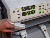
Her facility currently uses the Nicolet® SONARA® TCD system.  While these hospital-grade devices can be used for Emboli detection or interoperate scanning for PFO (Patent Foramen Ovale) closures, Ms. Torres' current assignments are primarily focused on neuro-monitoring for the detection of increased risks of vasospasms. As a seasoned technical expert in various levels of TCD field use, Ms. Torres shares field insight on the medical applications of this diagnostic protocol. "I see an opportunity to use TCD for patients with symptomatic carotid stenosis at our facility.”  


SPECIAL TRAINING
Her training on TCD imaging first started in 2001 with an intensive training course presented by Medical College of Georgia School of Medicine led by Dr. Robert J. Adams currently at the Medical University of South Carolina working directly on Sickle Cell patients. Dr. Adams is considered a pioneer in using transcranial doppler (TCD) and transcranial Doppler imaging (TCDi) to assess stroke risk in children with sickle cell anemia (SCA). He also helped discover that regular blood transfusions can reduce strokes by as much as 90 percent in these children.[4] Additional training came from Natus training experts who come from many areas such as the Swedish Institute of Neurology and a comprehensive text: Practical Guide to Transcranial Doppler Examinations by Dr. Andrei Alexandrov.


Maty Torres
, BHSc, RVT, Chief Vascular Technologist/Technical Director, Non-Invasive Vascular Lab.  Seasoned Vascular Technologist with a demonstrated history of working in the hospital and health care industry. Skilled in Vascular Sonography, Data Analytics, Health Informatics, and Peer Mentoring. Strong information technology professional with a B.H.Sc. focused in Health Care Administration from Nova Southeastern University, currently pursuing a Master’s in Business Administration from William Patterson University.


THE SCIENCE OF BLOOD FLOW AND DOPPLER TECHNOLOGY
Hemodynamics is defined as the study of blood flow in relation to the status of the circulatory system and homeostatic mechanisms of autoregulation.   Through the monitoring of blood flow, diagnostic analysis can provide many answers to the health and physiological status of the target area scanned as well as cell-level metabolism, the regulation of the pH, osmotic pressure and temperature of the whole body, and the protection from microbial and mechanical harm.[1]   Assessing injuries, inflammation or mutative growths (like cancer tumors), assessment of blood flow provides diagnostic answers about the severity of tissue disorders or tumor malignancy.

The expansion of ultrasound research and development broke new ground in 1982 when Dr. Rune Aaslid (Norwegian Neurosurgical researcher) first introduced Cerebral Hemodynamics with the implementation of Transcranial Doppler science to offer noninvasive transcranial recorded imaging of flow velocity in basal cerebral arteries.  His report detailed that placing an accurately configured Doppler ultrasound transducer in the temporal area (just above the zygomatic arch)offers quantitative data of the blood flow velocity in the middle, proximal anterior and posterior cerebral arteries.[1.2] This scan is critical for detecting vasospasms and for reviewing circulation in the brain for potential disease of the carotid and vertebral arteries. [1.5] 

In 1989, Dr. Aaslid published the first study on dynamic cerebral autoregulation in humans - a biological and metabolic function dedicated to stabilizing cerebral blood flow. TCD detects the slightest change in perfusion-- the volumetric measure at which blood is delivered to tissue, or volume of blood per unit time per unit tissue mass. Alongside the study of blood pressure, Cerebral Autoregulation is vital to maintaining life as it supports proper delivery of adequate oxygen (in the blood) and nutrients to the brain and the removal of CO2 & other waste products.[2]  As Dr Aaslid used the TCD by scanning through the transtemporal approach, others explored the intracranial arteries through the orbital (eye socket) window. By the early 90's, the next generation of development teams (Spencer, Seidel, Dobson & Moehring) improved on the Doppler innovation to detect microemboli and hemodynamic physiology. Today, TCD is widely accepted and utilized for an expanding set of clinical and research applications including  ischemic stroke, sickle cell disease, subarachnoid hemorrhage and vasospasm. 

According to cancer diagnostic imaging expert Dr. Robert Bard, the introduction of 4D Doppler technology aligned with the ultrasound's base design concept of providing instantaneous, real time readings.  "Let's take the case of breast cancer screening, the technician simply puts the probe on the breast, finds the area, pinpoints it, presses a button and seconds later you have the map showing the types of vessels, volumetric arterial density and spatial location of the vessels in relation to the adjacent organs. You have functions that give you a vessel density measurement which shows how aggressive this is. Instead of genetic markers (which are very popular), visually displaying tumor aggression  performed in a matter of seconds to show cancer vessel invasion is a game-changer for any early detection or monitoring facility. Tumor aggression by blood flow evaluation is used worldwide in nuclear medicine, CT scans and MRI technology, however, one of the simplest and most cost-effective alternatives is the non‐invasive 3D Doppler breast procedure."


INTRACRANIAL SCANNING WITH DOPPLER ULTRASOUND
In 1982, continued advancement led to the Transcranial Doppler (TCD) ultrasonography for outpatient and inpatient settings. By integrating the ability to study BLOOD FLOW into a low-frequency transducer, placing the probe on the temporal area measures the cerebral arteries to detect and quantify cerebrovascular activities, diseases and brain injuries.   Other applications include the diagnoses of vasospasm (VSP) after an aneurysm rupture, hemorrhage or hemodynamic changes after ischemic or cryptogenic stroke. It also enables the study of cranial pressure fluctuations.  TCD also offers significant clinical benefits in the monitoring of sickle-cell disease by scanning brain stem death and elevated raised intracranial pressure (ICP).  Added features allow for surgical and post-op monitoring of vasomotor functions as well as detecting critical disorders like a microembolism.

SUMMARY OF TARGET CONDITIONS FOR TCD APPLICATIONS 
•    Vasospasm /aneurysm
•    Sickle cell anemia, to determine a patient's stroke risk
•    Ischemic stroke
•    Intracranial stenosis/ blockage of the blood vessels
•    Cerebral microemboli


PART 2: VERIFYING SIGNS OF COVID-19 STROKE THROUGH IMAGING

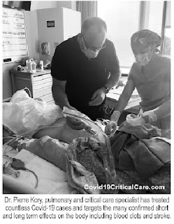
By: Robert L. Bard, MD  and Dr. Pierre Kory, MD

Early detection and prevention of arterial and venous disease is key to minimizing the effects of arterial obstruction & hemorrhage, brain aneurysms, and strokes from venous thrombosis.  The association of trauma to PTSD is now followed by advanced Doppler ultrasound and functional MRI. This abnormal physiology may also manifest as arterial dissection, collagen disease, inflammatory arthritis, dermatitis, ocular disorders, GI disturbances, limb pain, aneurysms of the brain and aorta. Devastating strokes in the Covid-19 era occur in the younger age group and the Latin population who are at higher risk.


INTRODUCTION
Interest in arteritis was elevated with the study of Tayakasu’s disease in the 1970's when advances in contrast arteriography diagnosed diffuse vascular involvement causing strokes and aneurysms in multiple sites. While this arterial inflammation is more common in Asians, in the US, blacks are nearly three times more likely to have a stroke at age 45 than whites. The pediatric population seems to be at higher risk for this arteritis as evidenced by their unusual rate of Covid-19 affliction affecting the vasculature and called “MULTIPLE ACUTE INFLAMMATORY SYNDROME“.  Birth control pills is a distinct cause of such disease in younger women while cancer, alcoholism and obesity raise the incidence at all ages. [3]

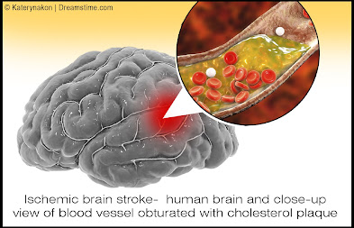
We have learned over the last century that blockages of coronary arteries to the heart and carotid arteries to the brain are precipitated by inflammation of the inner walls of the vessel, called the “intima”


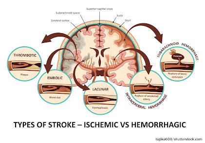
While thickening of the interior wall of vessels gradually occurs over time and is aggravated by diet, stress and hypertension (high blood pressure), the acutely disabling event occurs when there is an abrupt tear of the overlying plaque which ruptures debris which then forms a blood clot which blocks blood flow or the clot travels deeper into the brain and blocks blood flow. Similarly, abnormal heart rhythms such as “atrial fibrillation”, causes the pooling of blood in the heart which predisposes to clot formation and the clots can then travel into the brain causing a stroke. In Covid-19, the virus causing severe inflammation in the blood which then promotes clot formation which can travel through the vascular system and affect almost every organ system in the human body, with the brain and lungs being the most vulnerable. An article in September NEUROLOGY reported by Medscape documented the incidence of large artery stroke as the presenting symptom of COVId-19 was highest in men under the age of 50 years.  [4]

HISTORY
A medical research team at Metropolitan Hospital in New York first noticed unusual neurologic symptoms in young and middle aged patients in the late 1960s. As a division of the NY Medical College system, they were fortunate to have an active interventional radiology department specializing in neuroimaging and arteriography. The observation of distortion and occlusion of arteries supplying the brain, kidneys, GI tract and lower limbs to various degrees from single to multiple locations was closely linked to the Japanese disorder known as Tayakasu’s arteritis at the time and recently renamed “arteritis.” A clinical finding of this arterial inflammation in the abdominal aorta was pain in the upper abdomen by the great vessels by palpation. Astute physicians were successfully treating this with commonly available “aspirin.”

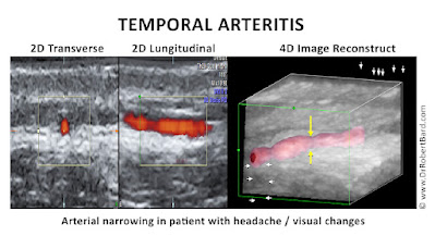
However, the chronic and diffuse nature of arteritis often weakened the vessel wall producing aneurysmal dilation and rupture. Today we find sophisticated non-invasive or minimally invasive modalities to be the first line of interrogation of vasculitis. [5]


COVID AND STROKE
COVID-19 was rapidly understood as a disease caused by severe and widespread inflammation and “hypercoagulability” (a tendency to spontaneously form clots in the blood vessels.) Autopsies have revealed extensive small vessel strokes, with such strokes often occurring despite aggressive blood thinner treatment and regardless of the timing of the disease course, suggesting that it plays a role very early in the disease process. In one autopsy series, there was a widespread presence of small clots with acute stroke observed in over 25%. In a recent review of the incidence of stroke in COVID-19, almost 2% of all hospital patients suffered a stroke, which is 8x higher than in patients with influenza. More worrisome is that this is almost definitely a gross underestimate given the many likely missed strokes in patients who died on ventilators who were too ill to obtain imaging, the general restrictions on and lack of autopsies, and the well-recognized decrease in the number of patients with acute stroke symptoms seeking medical attention in the COVID-19 era.  Another worrisome finding from a recent study of COVID-19 cases found that 45.5% of patients reported neurologic symptoms [6,7]. This under-recognized epidemic of neurological symptoms and strokes in COVId-19 highlights the need for more intensive imaging and investigation to achieve not only earlier recognition and improved treatment of patients but in furthering understanding of COVID-19 effects on brain function.

DIAGNOSIS BY IMAGING
Blood flow abnormalities in the arterial system are best study by Doppler imaging like the weather Doppler showing tornadoes. Multiple options exist for blood flow analysis including:

- Carotid Sonogram
- Carotid Doppler
- Eye Sonography
- Transorbital Doppler
- Contrast Enhanced Ultrasound
- Transcranial Doppler
- Hybrid Imaging
- 3D/4D Vessel Density Histogram
- Endoarterial 3D Doppler
- Retinal OCT
- Soft Tissue OCT
- Reflectance Confocal Microscopy
- Small Coil MRI
- 7 Tesla MRI


CAROTID SONOGRAM: While cerebrovascular disease is often diagnosed ex post facto after a catastrophic episode with MRI and CT, the non invasive Doppler analysis of the vascularity is generally checked with ultrasound for plaque and obstruction. A useful measure of the risk of coronary and cerebrovascular disorder is the carotid intimal thickness (CIMT). Standard depth of the inner wall thickness is a measure best obtained by high resolution sonograms since a reading over 0.9mm indicates increased risk. The newer sonogram units have depth resolution of 0.02mm making this a preferred non invasive option.


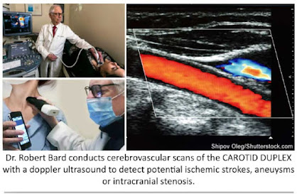
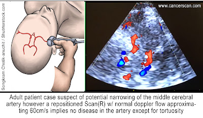
DOPPLER IMAGING TECHNOLOGIES

- COLOR DOPPLER - most common application where red is flow towards the probe and blue is flow away from the probe
- POWER DOPPLER - higher spatial resolution without directional flow correlation
3D POWER DOPPLER - allows volumetric analysis of vessel density used in treatment correlation where more vessels means increased neovascularity and fewer vessels correlates with clinical improvement
- ANGIODOPPLER – similar to color Doppler with higher spatial resolution
MICROVASCULAR DOPPLER-images capillary flow
B-FLOW DOPPLER-not true flow technology but observes motion of red blood cells directly

CAROTID DOPPLER: Flow abnormalities of turbulence and absence are commonly evaluated with this modality. Plaque forms more readily in aberrant flow patterns and high velocity regions accompanying narrowing.

EYE SONOGRAPHY: Sonofluoroscopy of the orbital soft tissues and eyes is performed in multiple scan planes with varying transducer configurations and frequencies. Power and color Doppler use angle 0 and PRF at 0.9 at optic nerve head. 3D imaging of optic nerve and carotid, central retinal arteries and superficial posterior ciliary arteries performed in erect position before and after verbal communication. Retinal arterial flow is measured. Optic nerve head bulging is checked as increased intracranial pressure may be demonstrable.

TRANSORBITAL DOPPLER: R/L ciliary arteries have normal Doppler flows of 10cm/s which is symmetric.

CONTRAST ENHANCED ULTRASOUND: Widely used European nonionic contrast injection allows imaging capillary size vessels and perfusion characteristics

TRANSCRANIAL DOPPLER: This measures the flow in the anterior, middle and posterior cerebral arteries as well as Circle of Willis.

3D/4D VESSEL DENSITY HISTOGRAM: Multiple image restoration and reconstruction shows retinal vessel density of 25% at the optic nerve head and adjacent region with quantitative accuracy.[8]

ENDOARTERIAL 3D DOPPLER: Microcatheters inserted into the arterial or venous system provide measurement of wall thickness and presence of inflammatory vessels inside the intima.

RETINAL OCT: Subtraction techniques done with OCT optical coherence tomography may show changes in the caliber of the retinal vessels with verbal ideation.

SOFT TISSUE OCT: The depth of penetration may be extended to 2-3mm allowing for analysis of vascular changes in erythematous or erythropoor dermal areas. Thrombosis may be observed.

REFLECTANCE CONFOCAL MICROSCOPY: This microscopic analysis of the cells also quantifies microvascular pathology and is a potential modality for studying vasculitis.

SMALL COIL MRI: High resolution systems used for animal study and superficial organs can image the intra-arterial anatomy including dynamic contrast imaging on standard 1.5T and 3T units.

7 TESLA MRI: High field systems analyze signal abnormalites rapidly with high resolution.

HYBRID IMAGING: Hybrid imaging refers to combining diagnostic modalities to assess disease and monitor therapy. 


Disclaimer: The information (including, but not limited to text, graphics, images and other material) contained in this article is for informational purposes only. No material on this site is intended to be a substitute for professional medical advice or scientific claims. Furthermore, any/all contributors (both medical and non-medical) featured in this article are presenting only ANECDOTAL findings pertaining to the effects and performance of the products/technologies being reviewed - and are not offering clinical data or medical recommendations in any way. Always seek the advice of your physician or other qualified health care provider with any questions you may have regarding a medical condition or treatment and before undertaking a new health care regimen, never disregard professional medical advice or delay in seeking it because of something you read on this page, article, blog or website.


REFERENCES

1) Hemodynamics: https://en.wikipedia.org/wiki/Hemodynamics

2) https://www.ncbi.nlm.nih.gov/pmc/articles/PMC5224659/

3). Hassani SN, Bard RL: Ultrasonic Diagnosis of  Aortic Aneurysms. J. Natl. Med. Assoc. 66:298-299, July 1974

4). Lande A, Bard RL: Arteriography of Pedunculated Splenic Cysts. Angiology 25:617-621, October 1974

5). Lande A, Bard RL, Rossi P: Takayasu's Arteritis: A World Entity. N.Y. State J. Med. 76:1477-1482, Sep 1976

6). Helbick Eur Radiol 30:5536-5538, 2020

7). Mao l  JAMA Neurol 2020 77:683-690

8). Bard RL , Gettz L, van der Bent S etal: Proceedings of the 4th Inflammatory Disease Summit at NY Academy of Medicine  Nov 2021


Tuesday, April 12, 2022

WHAT IS BIOFEEDBACK? PERSONALIZED SELF-REGULATING THERAPY

INTRODUCTION
IPHA's MedTech Reviews program is dedicated to the constant search for the most current advancements in health innovations.  We report, review, beta-test and challenge the efficacy of these inventions for public awareness of what works, and how they work. 

Here are just some of the latest non-invasive therapeutic technologies in our catalog of medical devices:
•  BIOLUMINESCENCE probes
•  RadioFrequency (RF) Therapy


Our next tour in our search for non-invasive health innovations is the science of BIOFEEDBACK—and we start our review with the internationally recognized pioneers from ONDAMED. Uniquely integrated with the features of PEMF/Pulsed Electromagnetic Field Therapeutics, this device offers localized tissue stimulation by induction of microcirculation within tissue. While the ONDAMED is approved by health authorities outside of the U.S. for pain relief, soft tissue injuries, and wound healing, the U.S. FDA regulates health claims to only include treatment for stress and stress related disorders.

An historically recognized electronic device earned its mark in progressive wellness, muscle relaxation and the alleviation of functional medical disorders (Jacobson/Harvard, 1938)- and evolved into the core of self-regulating therapeutics. In the early 1920's, a community of researchers founded the biometric readings as part of the early works of EEG, visceral learning & electromyography as the major functions of the biofeedback paradigm. EEG studies the basic principle that brain processes can be brought under voluntary control. Pursuit of the meditative, alpha dominant states continues to be part of today's EEG neurofeedback movement- supporting applications of brain wave control and the therapeutic signal of the device.[1]

In the 1970's the science of biofeedback dug its heels into the treatment of headaches and migraines, high blood pressure, urinary incontinence and lower back pain - among a list of other functional issues. Other devices and neurodiagnostics identify the use of biofeedback as an effective driver of self-regulating scanners of biodata.

- ELECTROMYOGRAPH (EMG): measures muscle fibers & neuromuscular studies. Neurofeedback may sometimes be used as a non-invasive treatment for ADHD, pain, addiction, anxiety, depression, and other disorders. [2]

- THERMAL FEEDBACK: reads migraine headaches & Raynaud's disease

- GALVANIC SKIN RESPONSE METER (GSR): measures electrical changes in the skin (sympathetic nervous arousal) supportive of psychotherapy & behavior therapy to track anxiety and cognitive/emotional threat response

According to Rolf Binder, chief engineer/inventor of the ONDAMED device and owner of the Ondamed Companies, and Dr. Silvia Binder, CEO of the Ondamed Companies, biofeedback is a mind-body process of electronic signals designed to send-then-receive (feedback) information about a person's physiological state, aimed at restoring control over involuntary bodily functions such as heart rate, muscle tension, blood flow, pain perception, and blood pressure. The (diagnostic) device sensors acquire feedback about specific aspects of your body. Once that information arrives, the goal of the device is to make subtle changes to the body that results in the desired effect. This might include relaxing certain muscles, slowing heart rate or respiration, or reducing feelings of pain. By doing this, people are often able to improve their physical, emotional, and mental health. For example, biofeedback can also be used to help people better manage the symptoms of a condition including insomnia, anxiety, depression, and pain.

The Association for Applied Psychophysiology and Biofeedback defines biofeedback as a process that allows people to alter their physiological activity in order to improve health or performance. Utilizing precise measurement instruments, information about the body's functions are provided to the user. [2]

BIOFEEDBACK THERAPY (e.g., ONDAMED) stimulates the body with gentle focused pulsed electromagnetic fields/PEMF in stressed areas that the biofeedback 'reports' to need help. This therapy is especially helpful to balance parasympathetic and sympathetic nervous systems, impacting the psyche, immune system, and endocrine system.


HOW DOES IT WORK: The biofeedback sensors "learn" about your specific physical signs and symptoms of stress and anxiety (ie. increased heart rate, body temperature, and muscle tension) through electrical sensors that are connected to specific areas of your body. During your session, your therapist will guide you through different mental exercises that may involve visualization, meditation, breathing, or relaxation techniques.  The ONDAMED Biofeedback System combines stimulation of gentle focused pulsed electromagnetic fields with Biofeedback. The feedback from the patient is done by monitoring the pulse rate and SpO2 displayed on the device. In addition, the practitioner palpates the patient’s radial pulse (aka vascular signal). This method allows personalizing stimulation of frequencies and sound along with placing applicators on most reactive stressed areas on the body for the patient’s fastest and lasting recovery.

"The ONDAMED device is an intelligent approach to providing stimulation with your body’s own communication language, which is electromagnetic. The brilliance of the ONDAMED method is the personalization of targeted localized frequency stimulation raising awareness to the patient to overcome stress and stress-related disorders."


"The ONDAMED device is an intelligent approach to providing stimulation with the body’s own communication language, which is electromagnetic. It’s not just going by symptoms or pathology or by what one knows about one frequency, but rather it is about combining a physical stimulation with biofeedback to personalize healthcare. Connecting with the patient by palpating the patient’s radial pulse while she/he receives ONDAMED’s therapeutic stimulation will give us the information as to what frequencies and where on the body these frequencies are needed most. It is overriding what we know or don’t know about the patient’s life health story. This method is comparable to a lie detector; we are accessing the individual’s innate wisdom by tapping into their communication pathways, and perhaps into part of their subconscious. Within minutes, the applicators can be placed on select areas for treatment. With biofeedback, we make the patient aware of what it is that they respond to—and where it is on the body that they need treatment. The patient’s autonomic nervous system is teaching the patient about their state of mind, their emotions, and biological consequences; this is all occurring while therapy is already running." 

- Dr. Silvia Binder


VIEWPOINTS

"As a PEMF medical protocol specialist, I’m experiencing the cellular response to PEMF with my patients every day. A dysfunction of the mitochondria can cause many disorders such as diabetes, Parkinson's disease and Alzheimer's. Research has shown that PEMFs recharge cells through ion exchange, bringing cellular energy back to its full potential and slowing the aging process of cells. Once cells are at their optimum potential, they are therefore able to operate more efficiently and for a longer period of time." - Joseph J. Toy / PEMF Medical Protocol Specialist.  (www.cliniquesneuroviesante.com)


NEW PROGRAM: Women's Diagnostic Network
Having access to the latest in compassionate experts and innovative diagnostic solutions for women is the first step to maintaining good health. Our alliance unites collaborative minds about the latest solutions from a wide range of modalities to offer optimum choices for all patients. Meet our current caregivers and educators for integrative care of women's chronic disorders. Together, we provide expansive research and info-sharing about all clinical options and protocols as well as testimonials and clinical viewpoints from active professionals in the field. 

The Women’s Diagnostic Network is a public informational resource backed by the Integrative Pain Healers' Alliance and the AngioFoundation. We are comprised of an all-volunteer collective of independent clinical experts, medical researchers and educators in specialized areas of clinical study. We support all areas of women's health interests including (but not limited to) CANCER CARE, ONCOLOGY, GYNECOLOGY, PHYSIATRY, DERMATOLOGY, NEUROLOGY, CARDIOLOGY & MENTAL HEALTH. Through clinical and academic collaboration, we unite to support improved patient evaluation, reporting on innovations and comprehensive diagnostic care. (visit WDN Webpage)

TOP HEADLINES
• 3/24 - Electromagnetic Breakthrough from Above our Northern Border: By Joseph J. Toy
• 3/16 - Essentials of SHOCKWAVE THERAPY: feat. Uran Berisha (IloveShockwave.com)
• 3/7 - Personalized Nutrition: Going beyond VitaminB12 for Vegan Diet Support: By: Dr. Bobbi Kline
• 3/7 - Integreative Medicine Review of Toxins
• 3/7 - The Sitting Culture: High Risk of Decline and Mortality: by Dr. Jonathan Kirschner
• 2/24 - Can PEMF Speed Up Healing Time? - by Dr. Jerry Dreessen
• 2/23 - Mental Health 101: Treating Incontinence Starts with Overcoming the STIGMA: by Dr. Bobbi Kline


REFERENCES:

1) Biofeedback, Mind-Body Medicine, and the Higher Limits of Human Nature:  https://www.aapb.org/i4a/pages/index.cfm?pageID=3383

2) https://www.ondamed.net/en/ondamed-biofeedback


Disclaimer & Copyright Notice: The materials provided on this website are copyrighted and the intellectual property of the publishers/producers (The NY Cancer Resource Alliance/IntermediaWorx inc., The AngioFoundation and Bard Diagnostic Research & Educational Programs). It is provided publicly strictly for informational purposes within non-commercial use and not for purposes of resale, distribution, public display or performance. Unless otherwise indicated on this web based page, sharing, re-posting, re-publishing of this work is strictly prohibited without due permission from the publishers.  Also, certain content may be licensed from third-parties. The licenses for some of this Content may contain additional terms. When such Content licenses contain additional terms, we will make these terms available to you on those pages (which his incorporated herein by reference).The publishers/producers of this site and its contents such as videos, graphics, text, and other materials published are not intended to be a substitute for professional medical advice, diagnosis, or treatment. For any questions you may have regarding a medical condition, please always seek the advice of your physician or a qualified health provider. Do not postpone or disregard any professional medical advice over something you may have seen or read on this website. If you think you may have a medical emergency, call your doctor or 9-1-1 immediately.  This website does not support, endorse or recommend any specific products, tests, physicians, procedures, treatment opinions or other information that may be mentioned on this site. Referencing any content or information seen or published in this website or shared by other visitors of this website is solely at your own risk. The publishers/producers of this Internet web site reserves the right, at its sole discretion, to modify, disable access to, or discontinue, temporarily or permanently, all or any part of this Internet web site or any information contained thereon without liability or notice to you.

Tuesday, February 15, 2022

A PEMF SUCCESS STORY: "GETTING MY LIFE BACK FROM CFS"

Interview by: Lennard Gettz, Ed.D & Roberta Kline, MD
Edited by: Dr. Robert L. Bard

Introduction

In support of personal leadership and proactive health, The Integrative Pain Healers Alliance applauds Ms. Suzanne Wheeler of Minneapolis, Minnesota as our Researcher of the Month.  After years of suffering a life-altering disorder that currently continues to challenge the scientific community of its root causes, Suzanne explored “outside the conventional box” of opioid prescriptions until she uncovered the one remarkable solution that got her back on her feet and joining life again.  Invoking CHANGE against all odds by diligently searching for what’s beyond the convenient takes courage and conviction.  It is this level of leadership that defines Alternative Health and Wellness to educate others about new answers that offer better results.

SHOT DOWN BY CFS
In 2017, I was diagnosed with Myalgic Encephalitis or Chronic Fatigue Syndrome. I had been essentially home bound for about two years and probably bed bound about 50% of my time. CFS was found to be the source of my 'all over body' pain- and so started my road to seeking out more targeted treatments. 

Life before CFS was very active. I worked in corporate America for 17 years as a regional manager for a large building facilities company and managed a team of about 3000 people. Prior to that, I was in the United States army for a decade. I was a Blackhawk pilot and a C 12 airplane pilot in the military before I entered corporate America. 

I started to feel significant pain after the birth of my 3rd daughter in Oct 2002.  I had a traumatic delivery due to a condition called placenta previa.  I lost over half my blood volume during the birth and received extensive blood transfusions.  I never felt the same after that experience.  About a week after delivery, the pain throughout my body started.  Over the years – it became worse and worse, and the fatigue became debilitating.  I kept pushing through with my job and family responsibilities which may have damaged my body even further.  


CFS is not an easy problem to resolve. I spent extensive time at Stanford University's chronic fatigue clinic with some of their top specialists.  I was also sent to the Workwell Foundation (CA) where I underwent a two day CPET (image insert) a cardiopulmonary exercise testing where they were able to determine what my anaerobic threshold was my VO2 max.  It was there that the clinicians found the extent of my metabolic dysfunction; my reports showed a VO2 reading equivalent to that of about a 95 year old. 

I had the kind of pain that radiated all over- throughout my spine, throughout my hips, legs, my knees, ankles, feet also throughout my arms. I had, peripheral neuropathy in my hands. I had trouble moving my hands and trouble typing and then also trouble walking. It developed to the point where in 2015 I was using a cane in my late forties to walk. I was desperate to get pain relief for more than a decade. I also had compression in my lower spine and my L4, L5, and also in my thoracic region (T 11 & 12) that caused extensive pain.  It wasn't an easy thing to figure out but through MRI, you can see the physical deformities- where imaging helped make everything pretty clear. But as to why it radiated through my body like it did, it took a lot of years to find out. 

DESPERATE FOR PAIN RELIEF
Since 2002, I tried everything- modalities from opioids to hip injections to spinal injections from a pain clinic. I saw chiropractors, I had acupuncture, tens units, electrical stim, laser treatments, extensive cranial psychotherapy, massage, a lot of ice and heat, a massage chair and a hot water Jacuzzi (which was the one thing that I responded to the best).

All pain relief from this point was temporary.  The base pain would eventually return and continue to get progressively worse. Years into this, it didn't seem like there was a lot left for me to try.  I learned that my condition was based on a specific virus that destroys your metabolic system and your body doesn't have the ability to oxygenate for energy. I was extremely tired all the time with post exertional malaise and my anaerobic thresholds became very low.  Our body builds up lactic acid with any exertion, with even minimal amounts of exertion. 

TAKING CHARGE: RESEARCH OUTSIDE THE BOX
In 2017, I learned about PEMF and its concepts of getting to the cellular level by helping my cells oxygenate.  Reading the many testimonials about it, it seemed to make better sense than the other modalities I tried so far.  User stories matched up with my symptoms and what I had been told by doctors was happening to my body. My sister introduced me to the idea upon seeing a presentation at a horse event, believe it or not. I became intrigued and acquired a rental and eventually purchased my  own model.

ALTERNATIVE HEALTH
I have HAD some wonderful physicians who are experts in their lane. At the height of my illness, I was seeing six different doctors who had me on so many medications (10 or 12 prescriptions) and I was still bed ridden- hence, nothing was working. 

Before I started using the PEMF, I had been essentially home bound for about two years and probably bed bound about 50% of my time. And if I was not in bed, I was seeking pain relief, usually in a hot bath. I would say within two weeks, the pain throughout my body had significantly subsided. I still had a little bit of joint pain but the overall soft muscle pain was GONE.  I had been taking opioids for over a decade prior, and I practically eliminated all that. It was, it was pretty unbelievable. Pain was a huge factor.  I wasn’t changing my doctors - but the one thing we all agreed on is that they found sudden improvement in me.  In my gut, I was confident of the changes were from the use of PEMF.


SUZANNE TODAY
I live a completely different life. To be clear, I'm not healed. I still have to live a more gentle life- certainly nothing like I used to do back in my army days or my early years working in corporate America- running and teaching aerobics and doing things like that. I still am not there to be able to do all that. The disease is still there, but the symptoms are managed and I have eliminated all the harsh, addictive medications. I am definitely getting so much more out of life without pain pills as opposed to being bed ridden and walking with a cane.

For anyone suffering like this, it's worth looking into. You want the expertise from the medical world, but you should blend the holistic treatments that are available- and then research it. If you search PEMF- read up on it! You'll find so many clinical studies and user testimonials that are coming up on that for all different ailments (not just mine) and they're seeing results. 

Thanks to PEMF, I'm able to participate with my family. I'm able to travel, go to the beach and take wonderful walks again. My sisters and I started my own business helping horses and people (with PEMF) be more pain-free and traveling again. It's hard not to be passionate about something that has that effect on your life. My husband says he got his wife back.


SPECIAL THANKS
The editors of this spotlight proudly gives thanks to Ms. Suzanne Wheeler for her generosity in sharing her story and her resources with us.  Additional thanks to Dr. Jerry Dreessen of the AOPP (Association of PEMF Professionals) and Pat Ziemer of Magnawave Inc. and Aura Wellness PEMF for coordinating our interviews, shared countless materials and conducted unending support to help our educational program bring new light to PEMF technology for chronic disorders and supportive testimonials in alternative therapeutics.   


ScienceNews Extra
Commentary on CFS/ME (Chronic Fatigue Syndrome and Myalgic Encephalomyelitis
By: Dr. Bobbi Kline

One of the key underlying findings in conditions causing chronic mental or physical fatigue is dysfunction of the mitochondria. Mitochondria are small structures located in the nucleus of every cell, and every cell contains thousands of them. These powerhouses produce the energy our bodies need to carry out every function in the form of ATP (adenosine triphosphate). When mitochondria don’t function at their best, or too many of them are destroyed, our energy levels suffer. While this can be due to inherited genetic disorders, most often it is seen as the result of chronic damage over time.

Many of the biochemical reactions in our bodies produce toxic versions of oxygen, hydrogen and nitrogen, including how we make ATP. These toxic molecules, which we call free radicals, have to be neutralized so they don’t damage the mitochondria. Our bodies have powerful antioxidant defenses to keep these in check. But when these protective systems become overwhelmed by too many free radicals, oxidative stress results and mitochondria are damaged.


IN THE NEWS

THE 2022 POST-MILITARY CRUSADERS COALITION 
2/11/2022- A 2022 initiative by community leaders launched the PMCC or Post Military Crusaders Coalition to launch an action plan for health resources for injured American veterans. Similar to the First Responders Cancer Resource project, this campaign supports all veteran advocates and service members support organizations by offering educational initiatives, alternative therapeutic modalities, sustainable diagnostic technologies and clinical research programs. 


CONTRIBUTORS

BOBBI KLINE, MD 
(Educational Dir. /Women's Diagnostic Group)
Dr. Kline is a board-certified ObGyn physician, Integrative Personalized Medicine expert, consultant, author, and educator whose mission is to change how we approach health and deliver healthcare. She helped to create the Integrative & Functional Medicine program for a family practice residency, has consulted with Sodexo to implement the first personalized nutrition menu for healthcare facilities, and serves as Education Director for several organizations including the Women’s Diagnostic Health Network, Mommies on a Mission. Learn more at https://bobbiklinemd.com 

 
ROBERT L. BARD, MD  (Diagnostic Imaging Specialist)
Having paved the way for the study of various cancers both clinically and academically, Dr. Robert Bard co-founded the 9/11 CancerScan program to bring additional diagnostic support to all first responders from Ground Zero. His main practice in midtown, NYC (Bard Diagnostic Imaging- www.CancerScan.com) uses the latest in digital Imaging technology has been also used to help guide biopsies and in many cases, even replicate much of the same reports of a clinical invasive biopsy. His most recent program is dedicated to the reporting of mental health diagnostic and innovative solutions including the use of modern neuromagnetic technologies and protocols in his MEDTECH REVIEWS program. 


Our friends and supporters:


Disclaimer: The information (including, but not limited to text, graphics, images and other material) contained in this article is for informational purposes only. No material on this site is intended to be a substitute for professional medical advice or scientific claims. Furthermore, any/all contributors (both medical and non-medical) featured in this article are presenting only ANECDOTAL findings pertaining to the effects and performance of the products/technologies being reviewed - and are not offering clinical data or medical recommendations in any way. Always seek the advice of your physician or other qualified health care provider with any questions you may have regarding a medical condition or treatment and before undertaking a new health care regimen, never disregard professional medical advice or delay in seeking it because of something you read on this page, article, blog or website.

Monday, February 7, 2022

PEDIATRIC MYOCARDITIS & NATIONAL DETECTION MOVEMENT (Part 1)

Written by: Dr. Robert L. Bard

Since the advent of Covid-19 Long Haul studies in 2021, the medical diagnostic community shifted into overdrive- seeking out all available screening and examination protocols to assess health problems called POST-ACUTE SEQUELAE (PASC). One of the recent Covid-19 related headliners is the rise in cases of MYOCARDITIS in children 16 years and under. CDC Reports link the pathological impact with covid infection since it is proven that Viral infections are a common cause of myocarditis. 

Between early 2020–2021, patients with Covid-19 had nearly 16 times the risk for myocarditis[1]. According to the CDC, in a study of myocarditis cases, 2,116 (41.7%) had a history of Covid-19.  In addition, cases of myocarditis reported to the Vaccine Adverse Event Reporting System (VAERS) indicated links between Myocarditis and Pericarditis to come from the mRNA Covid-19 vaccination (especially in male adolescents and young adults) more often after the second dose.[2].

Myocarditis is defined as an inflammatory disorder of the heart muscle (myocardium) leading to cardiac dysfunction. It is also recognized as myocardial cell death [3]. Checkups for this also reviews for PERICARDITIS (the inflammation of the outer lining of the heart). Various causes of myocarditis includes: Viral Infectious including adenoviruses, echoviruses, enteroviruses like the coxsackie viruses. In addition, predisposition can occur from those with Autoimmune diseases such as Celiac disease, Churg-Strauss syndrome, Crohn disease, Kawasaki disease, lupus, rheumatoid arthritis, sarcoidosis etc. (See NIH chart for full list of causes- [4])


STANDARDIZED DIAGNOSTICS

As with any critical disorder, detecting early stages of myocarditis allows for a higher opportunity to treat and even eliminate the health risk.  In children, symptoms include: Fever, Fainting, Breathing difficulties, Rapid breathing, Chest pain and Rapid or irregular heart rhythms [5]. In adults, symptoms range from chest pain, shortness of breath, at rest or during activity and fluid buildup with swelling of the legs, ankles and feet. To prevent possible heart damage, a cardiologist may order one of a number of imaging options:

▪ Electrocardiogram (ECG or EKG)
▪ Chest X-Ray
▪ Heart MRI
▪ Blood Tests
▪ Doppler Ultrasound for Acute Myocarditis
▪ Cardiovascular MR Elastography (MRE)
▪ Ultrasound Elastography

See expanded details on diagnostic protocols, visit:  http://pediatricscan.com/myocarditis.html


MedNews Extra
Saving Lives Through Advocacy & Research:
FOR ELYSA FOUNDATION

Around the first week of Feb, 2022, our clinical diagnostic researcher, Dr. Robert Bard launched his PediatricScan.com 2.0 in NYC‐ which included a Pilot program for Myocarditis Screening through the use of advanced Doppler Ultrasound Imaging.  To establish the clinical network for this program is to connect with ICU specialists & Cardiologists as well as all associations supporting Myocarditis research.

We met the directors of a remarkable national advocacy foundation called FOR ELYSA FOUNDATION‐ a non‐profit organization dedicated to promoting Education, providing Light, and supporting Research in the areas of viral myocarditis and pediatric sudden cardiac arrest. (www.ForElysa.org). Mrs. Jana Rojas and husband Jaime Rojas from Kansas City developed this organization inspired by the loss of her vibrant little girl,  Elysa Louise Rojas who passed away at the tender age of two years old. " In Elysa’s case, a common childhood virus was responsible for her myocarditis. The virus either attacked Elysa’s heart directly or caused her immune system to attack her heart muscle in a “friendly fire” fashion while trying to fight the virus. The inflammation in her heart increased drastically and very quickly to the point of sudden cardiac arrest. Doctors and scientists do not fully understand the mechanisms within the body that cause a virus to “go haywire” in the immune systems of individuals with myocarditis. There is currently no way to predict when/if this will occur."


The FOR ELYSA FOUNDATION is one of our first advocacy friends in pursuit of bringing national awareness and supportive clinical research for myocarditis diagnostics and prevention. According to the ForeElysa.org website, Myocarditis is a disease marked by inflammation and damage of the heart muscle. There are many causes of myocarditis, including viral infections, autoimmune diseases, environmental toxins, and adverse reactions to medications. The most common cause of myocarditis in North America is viral infections. Myocarditis usually attacks otherwise healthy people. It is believed that 5 to 20% of all cases of sudden death in young adults are due to myocarditis. Although the exact incidence of myocarditis is not known, it is estimated that approximately 343,000 people die of myocarditis and its major complication, cardiomyopathy, each year. The prognosis is variable but chronic heart failure is the major long term complication. Myocarditis and the associated disorder of idiopathic dilated cardiomyopathy are the cause of approximately 45% of heart transplants in the United States.  

Materials in this excerpt are published with express consent from The For Elysa Foundation.  For complete Information, visit www.FORELYSA.org


HOW & WHEN TO CHECK FOR PEDIATRIC MYOCARDITIS
By: Jana Rojas
The tricky thing with myocarditis being virally mediated is that Elysa could have had a heart scan a week before she died (the day before she contracted the virus that wrecked her heart), and it would have been normal. I am hesitant to insinuate that imaging could "clear" a patient and provide a clean bill of health without noting that this can and does occur spontaneously after viral infections, and so testing while ill or post-virally is actually the key message and window of opportunity for myocarditis detection.
In my mind, the primary role for cardiac diagnostic imaging as it related to myocarditis specifically would be for: 
1) acutely ill children in ED/urgent care/hospital inpatient settings
2) children exhibiting the signs and symptoms you have outlined (fainting, sudden fatigue, shortness of breath, chest pain, palpitations), 
3) after known Covid or other viral infection with prolonged or delayed healing (ie ongoing fatigue, shortness of breath, etc)  
4) and possibly PRE-PARTICIPATION SPORTS PHYSICALS. The pre participation screenings would be enhanced cardiac screenings in general to ideally pick up congenital heart defects and other concerns as well as myocarditis. 


TROUBLESHOOTING MYOCARDITIS
By Bobbi Kline, MD (Integrative Physician / Genomic Research Specialist)

As a mom, my heart grieves for parents, including Elysa’s, who suffer such devastating tragedy. It’s the worst thing you hope never happens to your child, and I truly admire parents who turn a tragedy into something positive. It requires such amazing strength, courage and grace. As a physician, I find my self immediately asking "Why do these things happen? How can we predict or prevent them?" As clinicians, we look for patterns to help guide diagnosis and treatment. We know what to expect, but sometimes they can lull us into a false sense of security. Childhood viruses, as any parent knows, are an expected part of those early years. 
But what happens when they turn out to be something more? That’s where pattern recognition is crucial. When something obviously falls outside those patterns, it’s a signal to question and go deeper. But what happens when you don’t even recognize that deviation? What if something is so uncommon or so subtle that it’s hard to detect among all the noise? Post-viral myocarditis is one of those conditions, and I’m glad to see it now in the spotlight. Raising awareness is a key first step. While COVID-19 has certainly helped to highlight this condition, it goes further than COVID. Many common childhood viruses have been implicated in causing myocarditis, but most people are completely unaware. I admit that it was not something I ever really thought about as my kids were growing up. And I am not alone. Educating clinicians as well as parents on what to look for, when to be alarmed, when to go deeper is crucial. This alone will save lives. 


But it’s only the first step. We also need better tests and tools to quickly and easily identify who is at risk, and better treatments for helping these children. This requires a multidisciplinary approach that includes better diagnostics including noninvasive technology, along with effective medications and other treatments. It also includes the burgeoning field of genomics and personalized medicine, both to provide a better understanding of the why, as well as a powerful tool to predict and prevent. For, at the heart of this, is understanding each child’s uniqueness in a way that empowers.  Two studies have been published this year that have the potential to leverage the power of DNA to identify who is at risk for developing myocarditis after a viral infection. Not only that, but also which of those children are most likely to recover, and therefore need fewer interventions, and which of those children are most at risk for sudden death and require much more intensive treatment and support. And, in today’s world, we also need the power of legislation to make sure everyone has access to this higher level of care. There is much promise to change the trajectory of this devastating illness, and it is only through advocacy such as this that it will happen. 


1) Morbidity and Mortality Weekly Report (MMWR): Association Between Covid-19 and Myocarditis Using Hospital-based Admin Data 3/2020-1/2021) https://www.cdc.gov/mmwr/volumes/70/wr/mm7035e5.htm
3) MR Imaging of Myocardial Infarction | RSNA-Radiological Society of North America  / https://pubs.rsna.org/doi/10.1148/rg.335125722
4) The Diagnostic and Clinical Approach to Pediatric Myocarditis: A Review of the Current Literature (NCBI/NIH)  https://www.ncbi.nlm.nih.gov/pmc/articles/PMC6352488/
5) Diagnosis and Management of Myocarditis in Children (American Coll. of Cardiology) https://www.acc.org/latest-in-cardiology/ten-points-to-remember/2021/07/09/17/31/diagnosis-and-management-of-myocarditis

Disclaimer: The information (including, but not limited to text, graphics, images and other material) contained in this article is for informational purposes only. No material on this site is intended to be a substitute for professional medical advice or scientific claims. Furthermore, any/all contributors (both medical and non-medical) featured in this article are presenting only ANECDOTAL findings pertaining to the effects and performance of the products/technologies being reviewed - and are not offering clinical data or medical recommendations in any way. Always seek the advice of your physician or other qualified health care provider with any questions you may have regarding a medical condition or treatment and before undertaking a new health care regimen, never disregard professional medical advice or delay in seeking it because of something you read on this page, article, blog or website. None of the information provided should be interpreted to be or is meant to be medical advice, suggestions, or counseling.